
The Human BioMolecular Atlas Program (HuBMAP)
The Human BioMolecular Atlas Program (HuBMAP)
Image of the Week
Some of the most amazing things to come out of the HuBMAP Consortium are the images of healthy human tissues generated by our researchers.
Here, we collected them in one place to celebrate the work of these talented individuals.
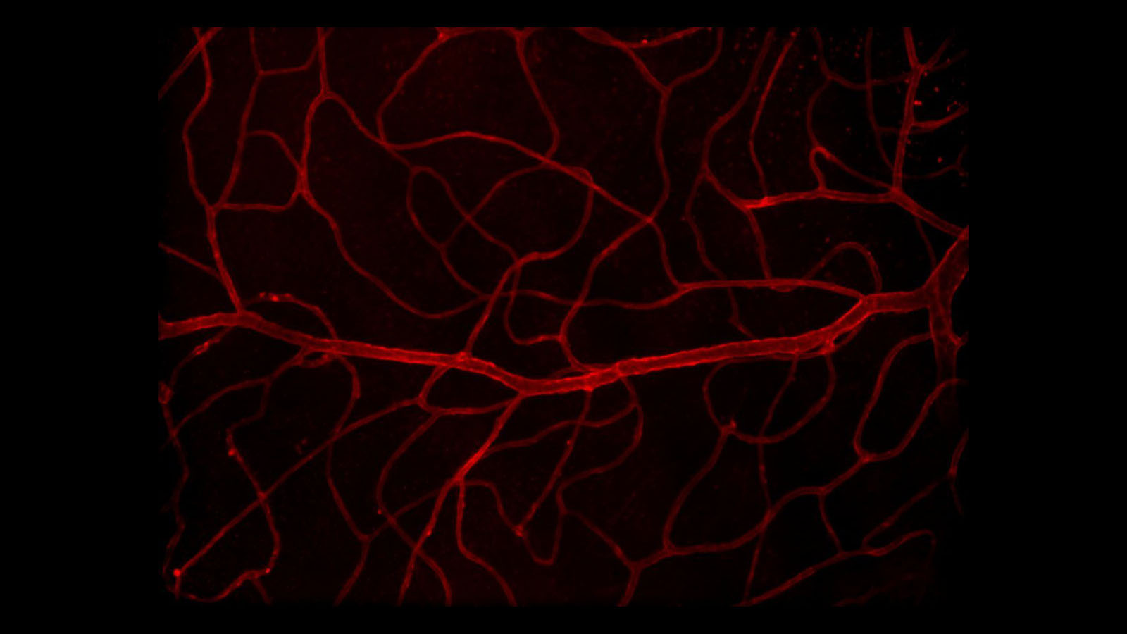
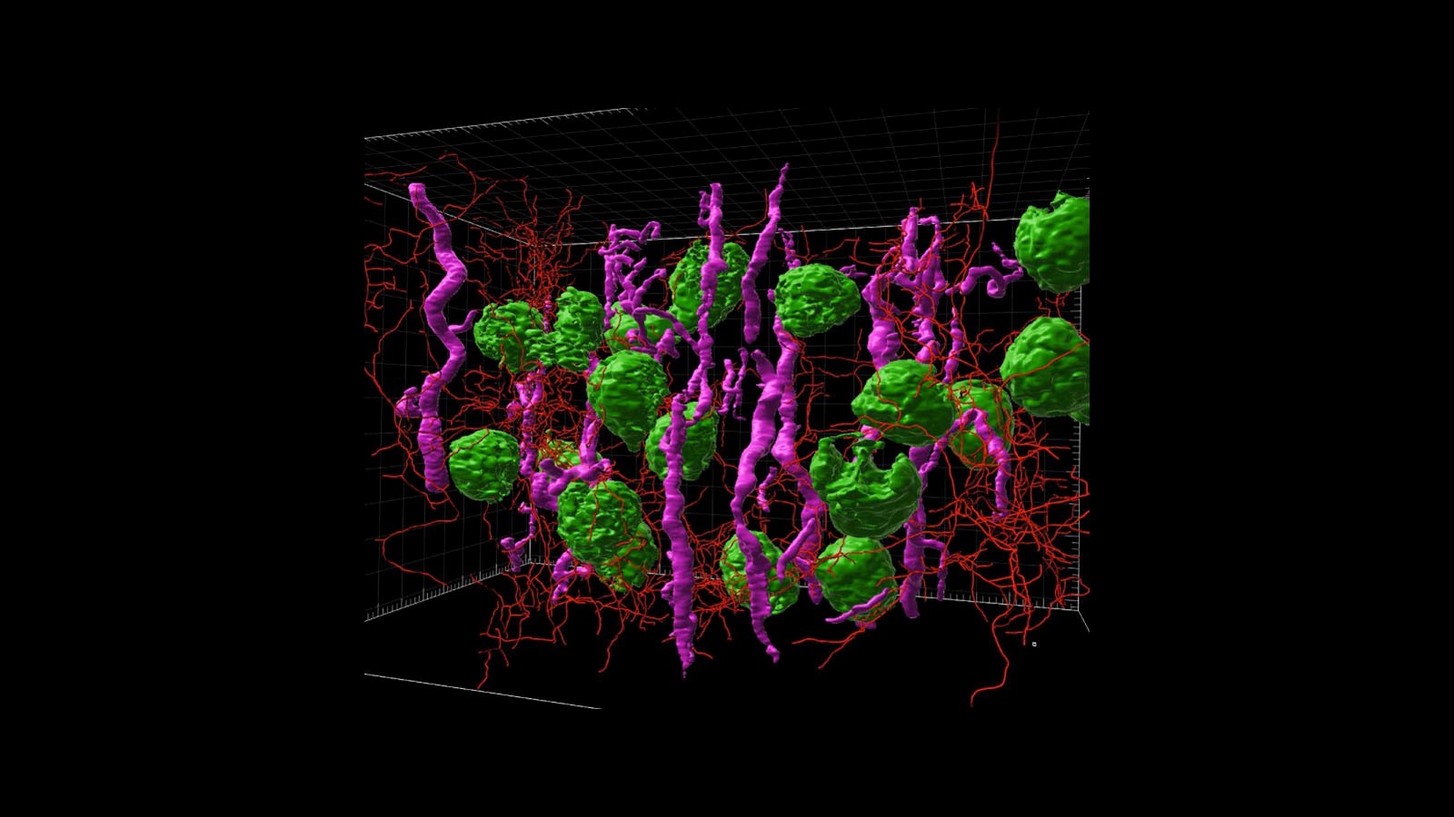
3D microscopy image of vasculature in human retina courtesy of Dr. Angela Kruse of the Spraggins Lab at Vanderbilt University
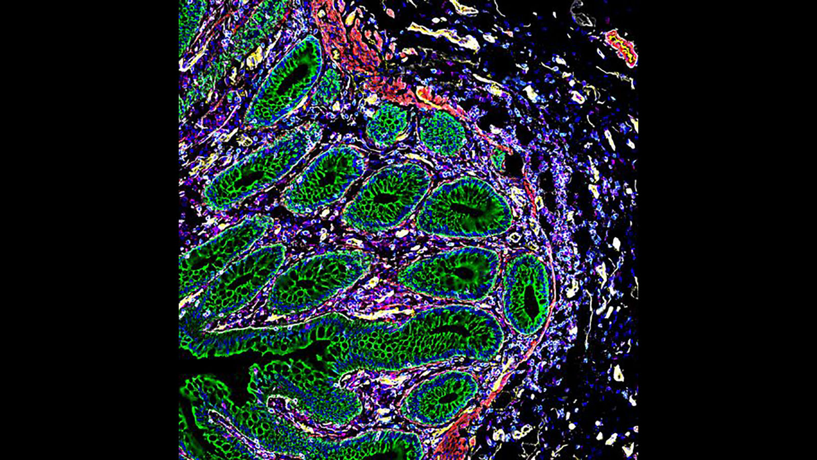
#CODEX image of healthy human intestine courtesy of Dr. John Hickey at the Nolan lab at Stanford University
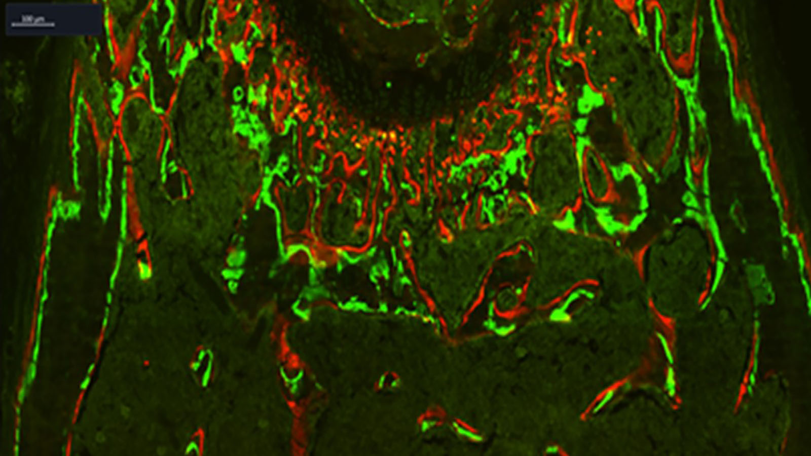
Image of a femur made with fluorescent dyes courtesy of Dr. Peter Maye at UConn.
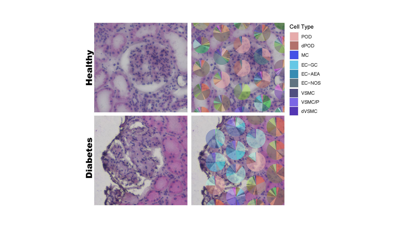
Spatial transcriptomics image of both healthy and diabetic kidneys from Dr. Sanjay Jain's lab at Washington University in St. Louis
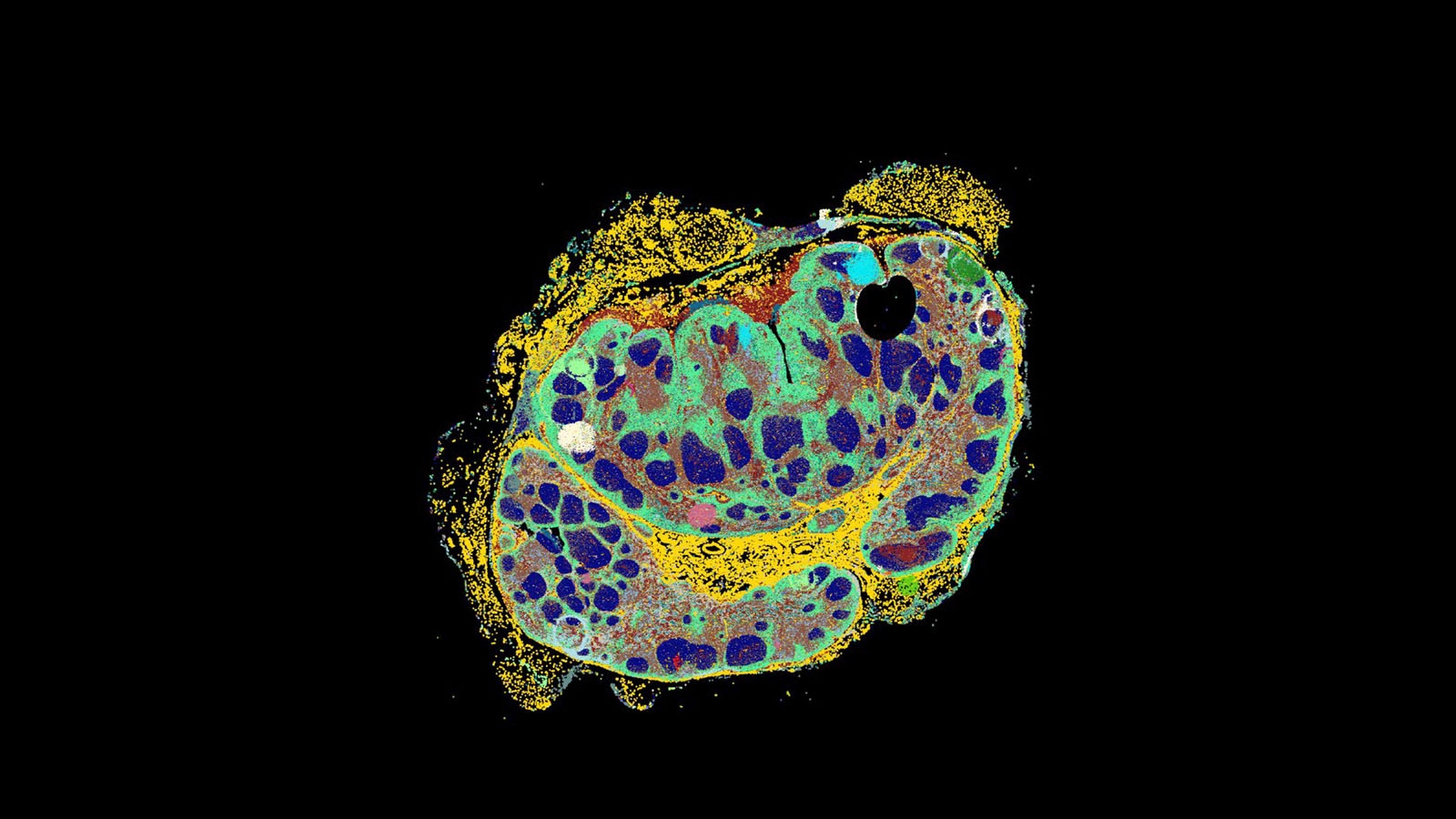
CODEX image of a lymph node from Archie Enninful at Rong Fan's lab at Yale
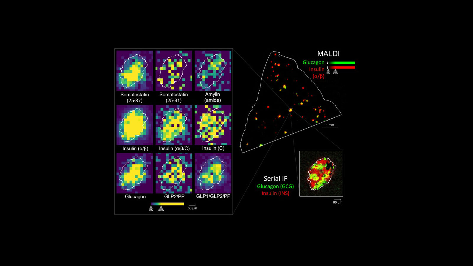
MALDI and serial immunofluorescent images of human pancreas from Dr. Kevin Zemaitis at PNNL
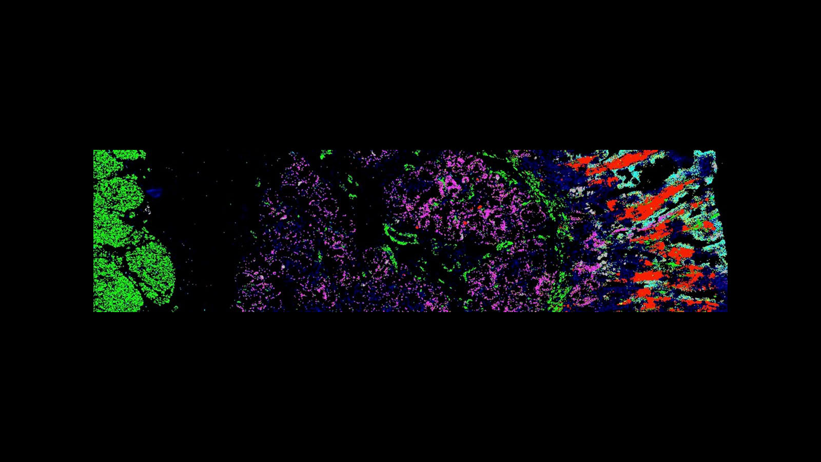
Molecular Cartography image of RNA within polyps from familial adenomatous polyposis patients, courtesy of Dr. Chenchen Zhu at the Snyder Lab at Stanford University
3D rendered light sheet microscopy image from human kidney cortex courtesy of Drs. Praveen Krishnamoorthy, Bo Zhang & Sanjay Jain at Washington University of St. Louis .
CODEX image of human intestine from Dr. John Hickey at Stanford University
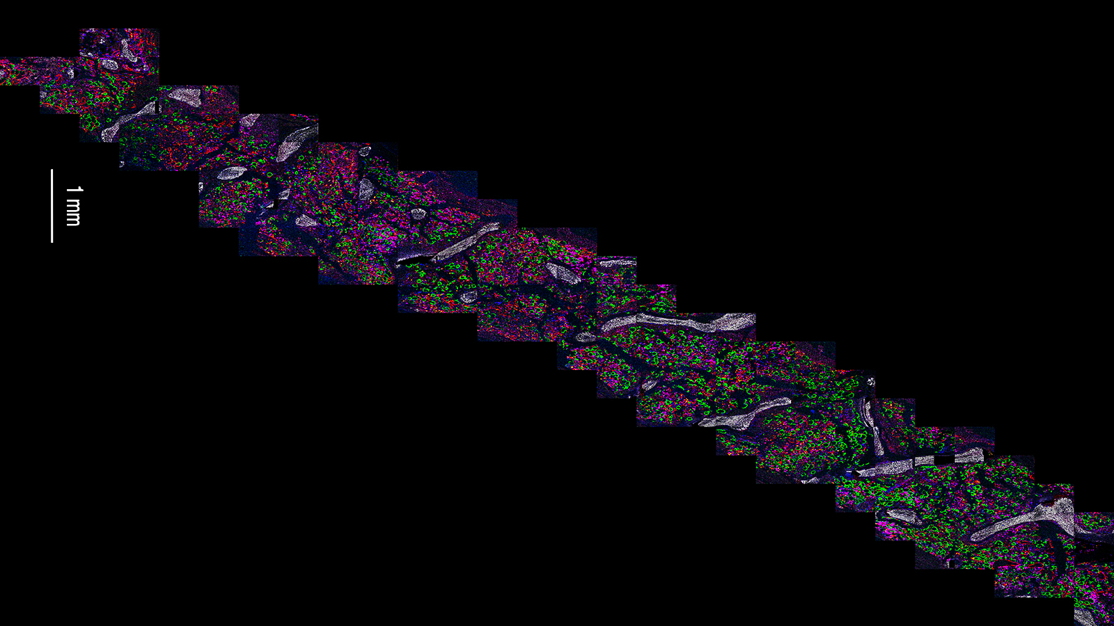
MIBI-TOF image series of healthy human bone marrow core, courtesy of Dr. Patricia Favaro from the Bendall lab at Stanford University

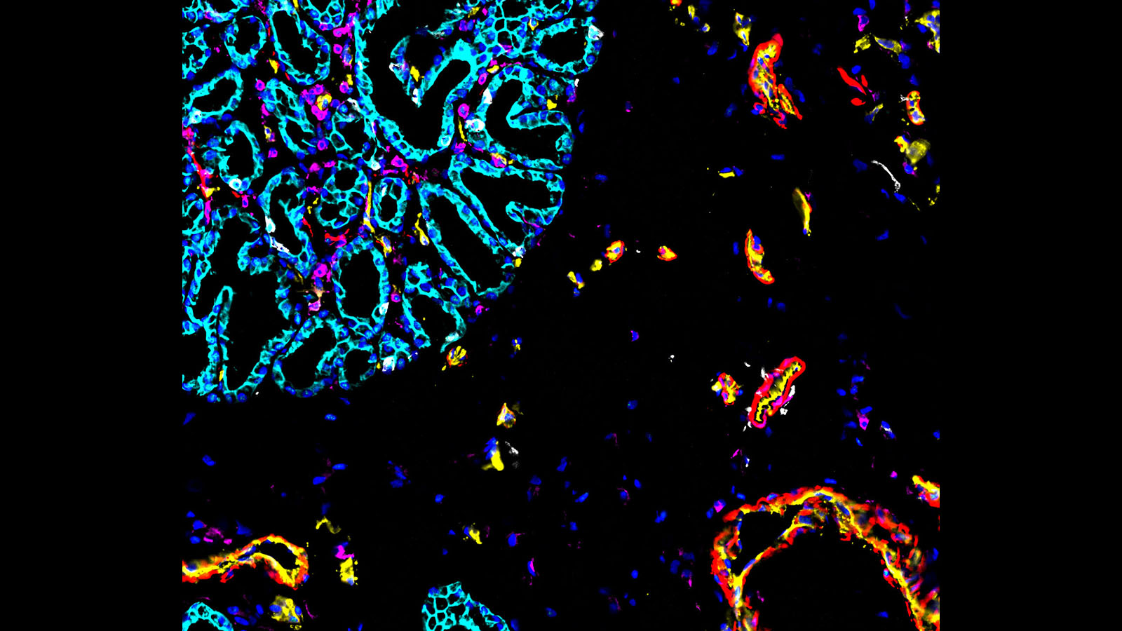
CODEX image of healthy human kidney, courtesy of Drs. Tarek Ashkar (Indiana University) and Sanjay Jain (Washington University at St. Louis)
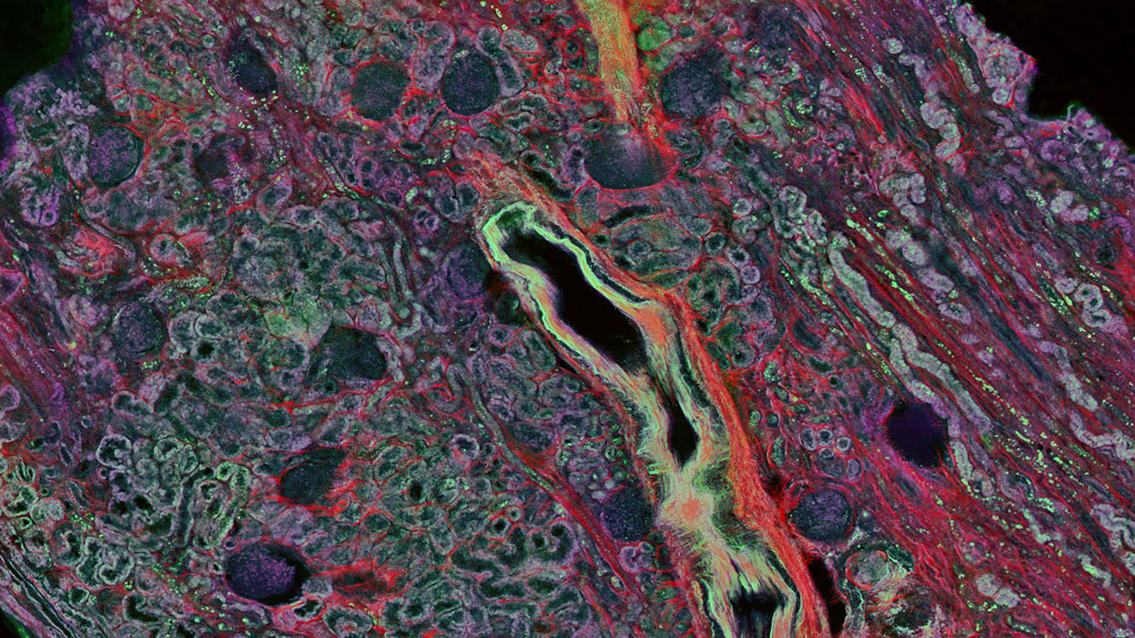
Autofluorescence image of human kidney cortex from Anthony Fung @UCSD showing the variations in cellular metabolism
