
The Human BioMolecular Atlas Program (HuBMAP)
The Human BioMolecular Atlas Program (HuBMAP)
Image of the Week
Some of the most amazing things to come out of the HuBMAP Consortium are the images of healthy human tissues generated by our researchers.
Here, we collected them in one place to celebrate the work of these talented individuals.
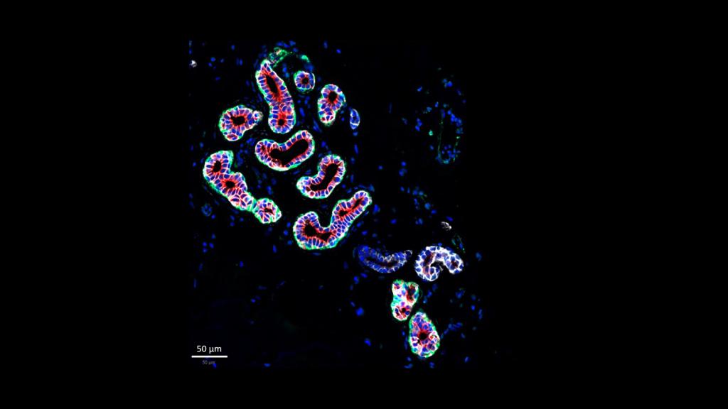
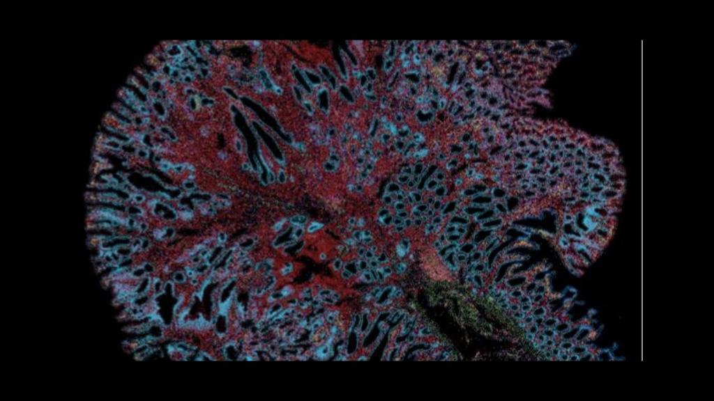
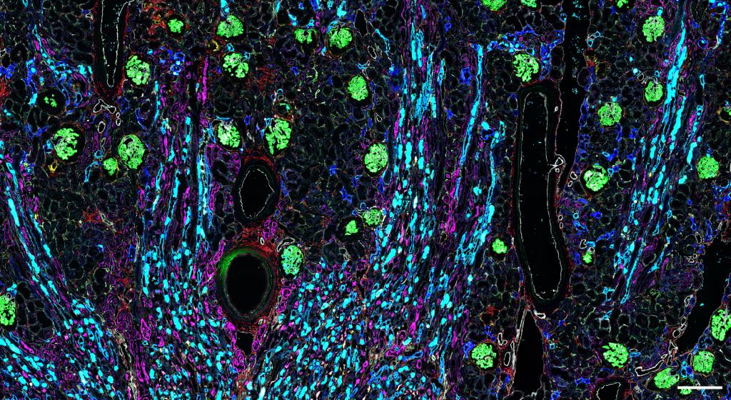
CellDIVE image of glands in skin, courtesy of Dr. Liz McDonough from GE
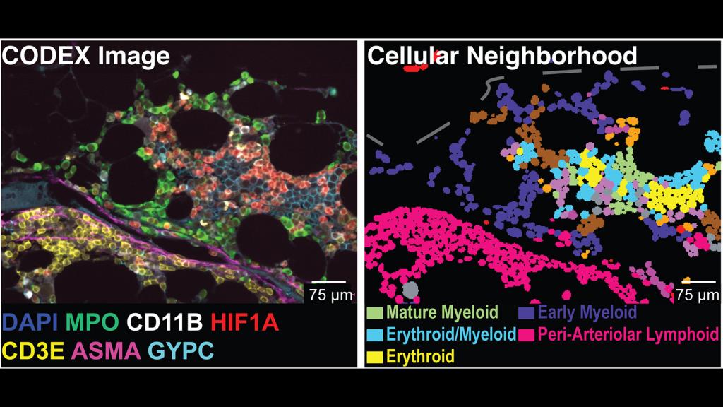
Left panel - CODEX of bone marrow, right panel - visualization of neighborhoods within the bone marrow sample, courtesy of Kai Tan's lab at UPENN
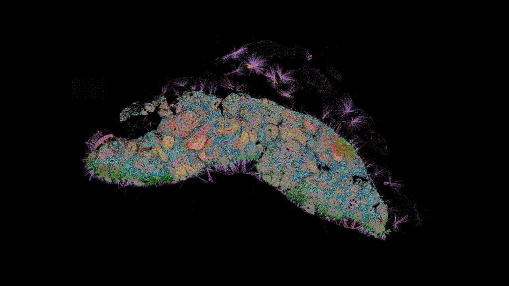
CODEX image of a lymph node, courtesy of Archie Enninful of Yale
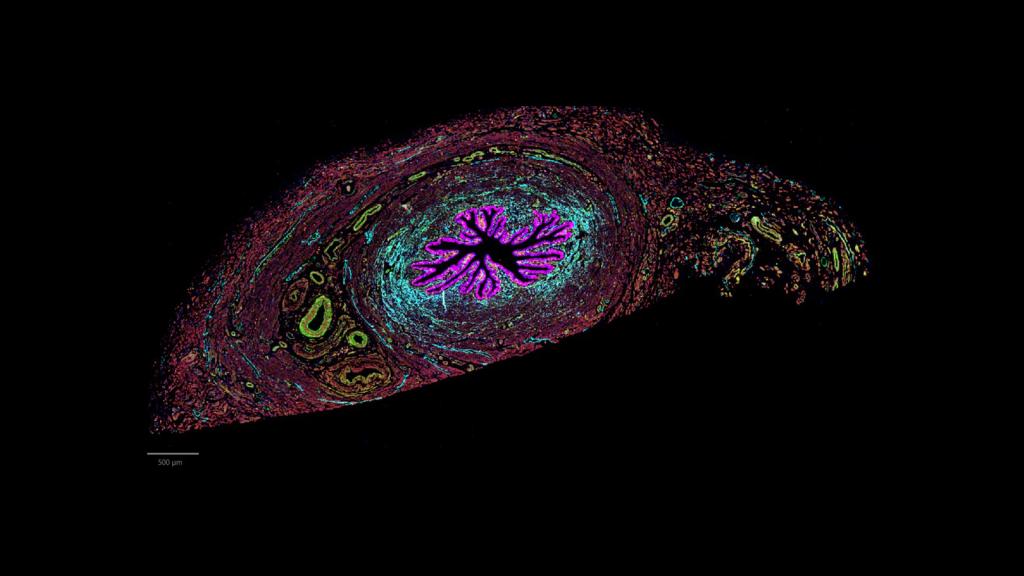
CODEX image of the isthmus, the connection between the uterus and Fallopian tube, courtesy of Dr. Kate O'Neill at UPENN
Xenium image of intestines courtesy of Dr. Chenchen Zhu of Mike Snyder's lab at Stanford University
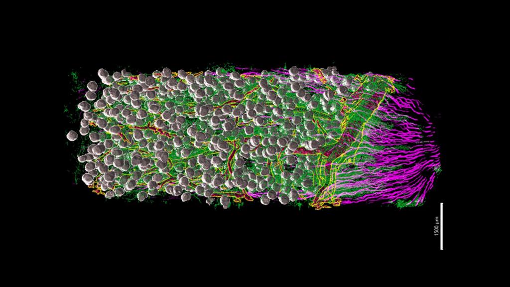
Lightsheet Microscopy image of kidney courtesy of Liam McLaughlin in Dr. Sanjay Jain's lab at WUSTL
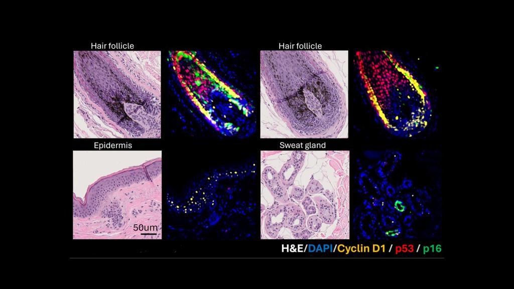
H&E and MxIF images of hair follicles, sweat gland, and epidermis in human skin, courtesy of Dr. Pei-Hsun Wu at JHU.
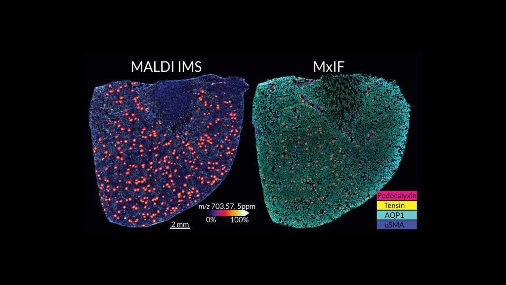
MALDI and MxIF images of healthy kidney from Allison Esselman of Vanderbilt University
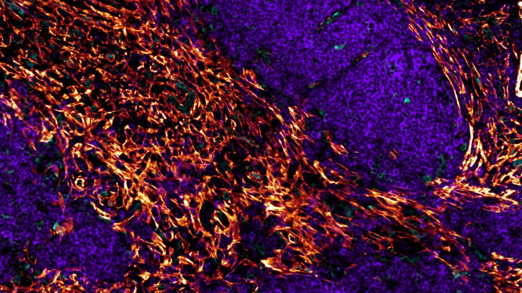
CODEX image of healthy kidney from Dr. Tarek Ashkar of Indiana University
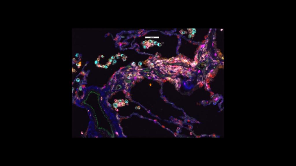
CellDIVE image of healthy lung, courtesy of Dr. Gloria Pryhuber at URMC
IBEX image of lymph nodes courtesy of Dr. Andrea Radtke in the Germain Lab at NIAID
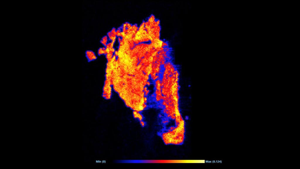
DESI-MSI image of linoleic acid throughout the left ventricle of the heart, courtesy of Taruna Neelakantan at Columbia University.
