
The Human BioMolecular Atlas Program (HuBMAP)
The Human BioMolecular Atlas Program (HuBMAP)
Image of the Week
Some of the most amazing things to come out of the HuBMAP Consortium are the images of healthy human tissues generated by our researchers.
Here, we collected them in one place to celebrate the work of these talented individuals.
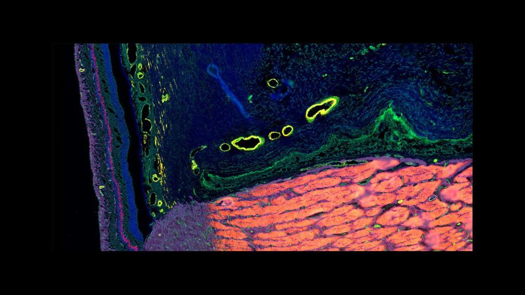
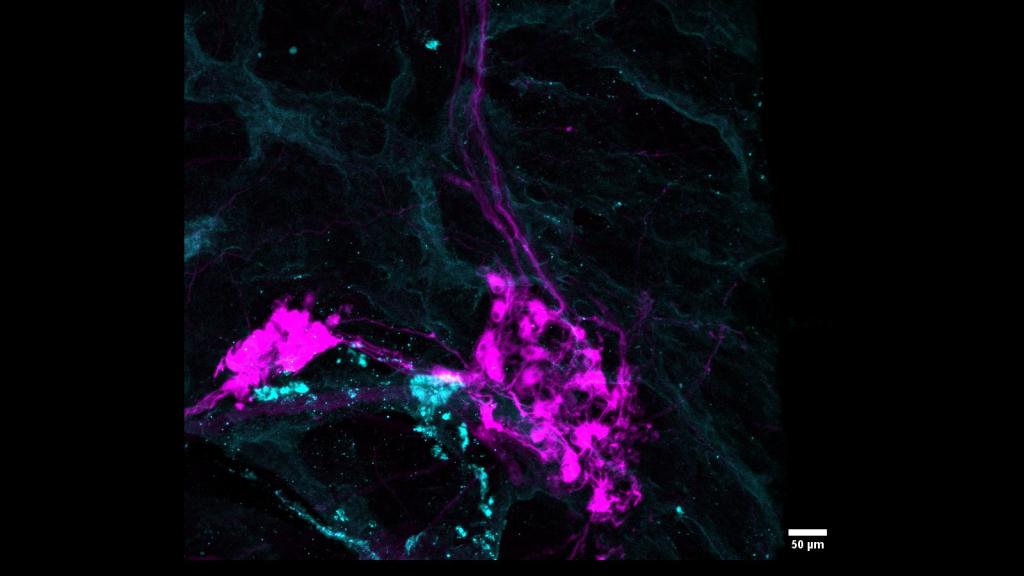
CODEX image of a human optic nerve courtesy of Dr. Angela Kruse, at Spraggins Lab at Vanderbilt University
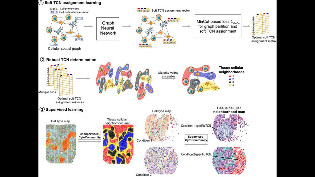
Infographic of CytoCommunity, a new algorithm developed by Dr. Kai Tan at CHOP that identifies types of cells neighborhoods
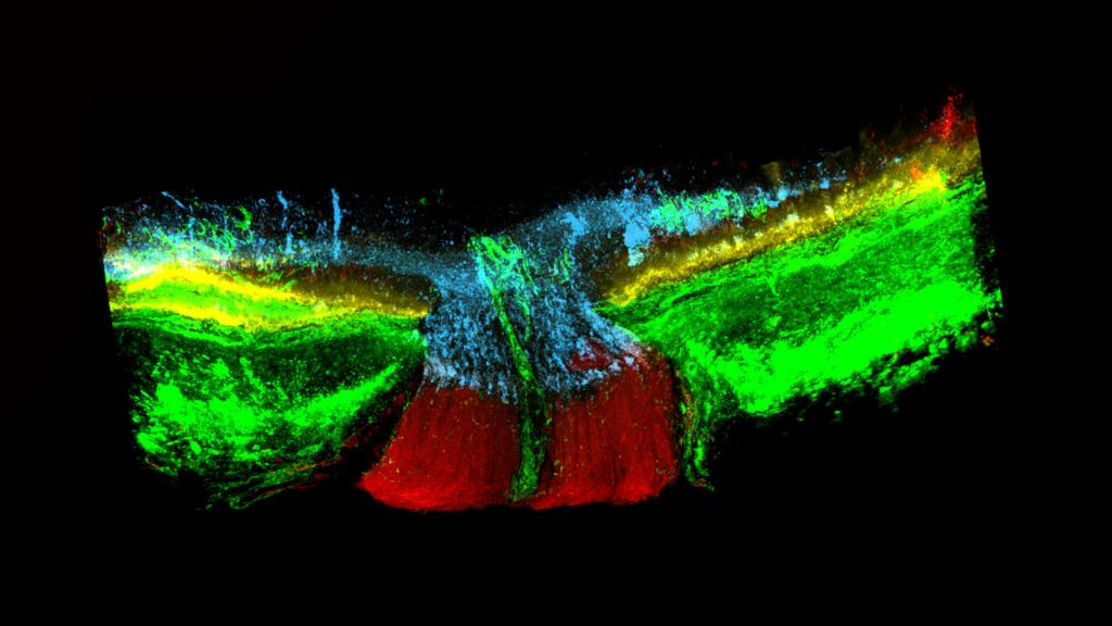
MALDI IMS image courtesy of Dr. David Anderson at Vanderbilt University of the lipids within the optic nerve
MALDI-MSI image, courtesy of Dr. Brittney Gorman, at Pacific Northwest National Laboratory, of the lipids within the lung
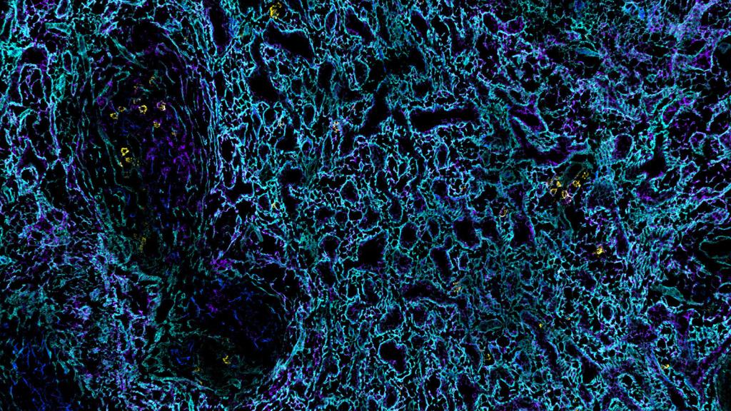
Phenocycler Fusion image by Drs. Gloria Pryhuber and Jeffery Purkerson, at the University of Rochester Medical Center, of the surface of the lung
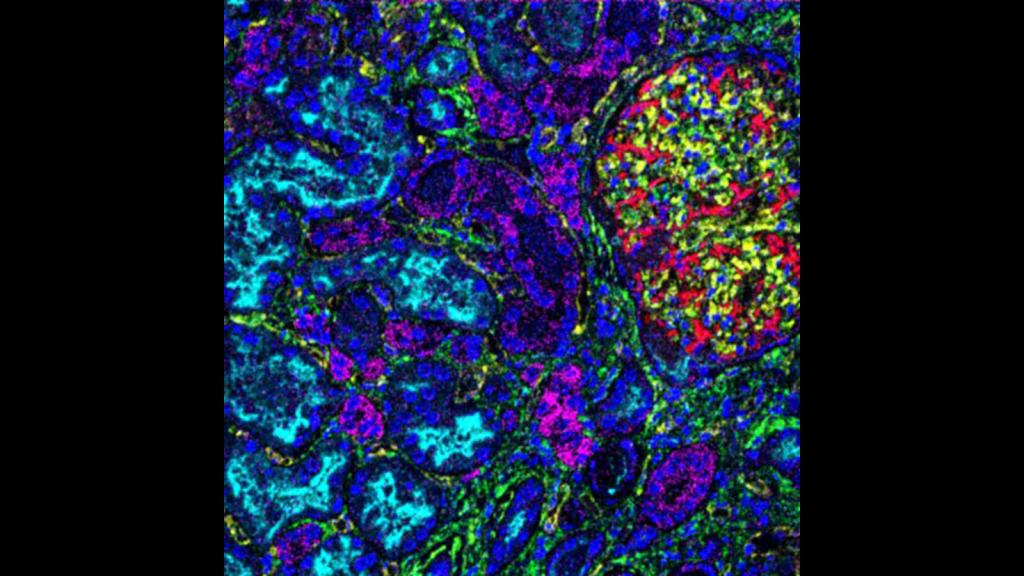
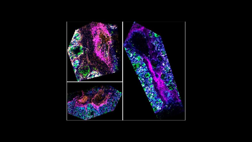
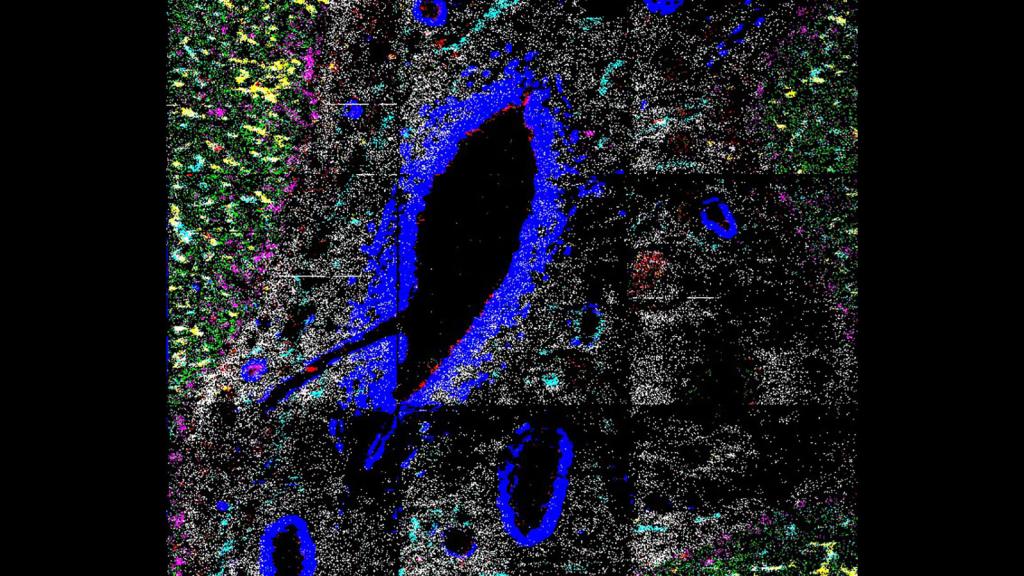
MIBI-TOF image by Dr. Albert Tsai at the Bendall Lab at Stanford University of a glomerulus and surrounding tubules within a human kidney
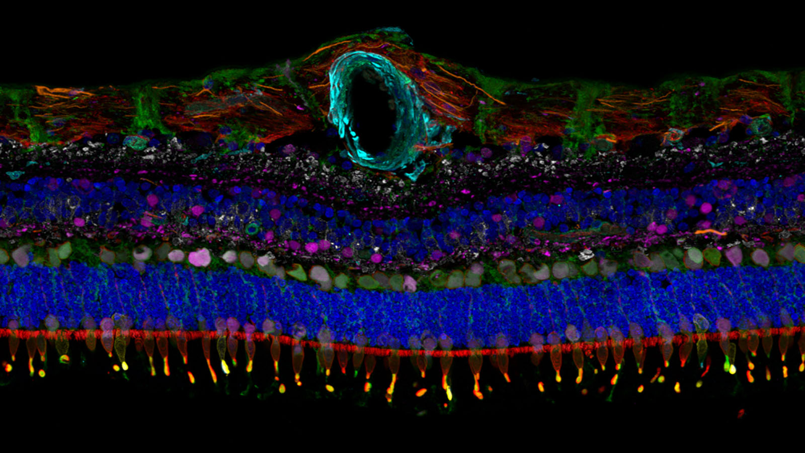
IBEX image of a human retina from Dr. Colin Chu at UCL Institute of Ophthalmology
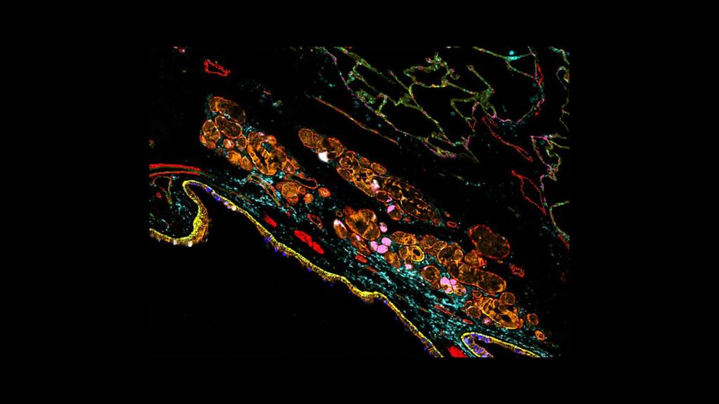
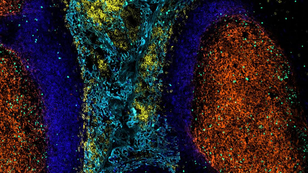
CODEX image of a neural node in the pancreas, courtsey of Dr. Angela Kruse in the Spraggins lab at Vanderbilt University
This is an IBEX image of a human spleen made by Dr. Andrea Radtke of the Germain lab at NIAID
This is a MASCima image of a human tonsil, made by Drs. Werner Muller & Andreas Bosio at Miltenyi Biotec
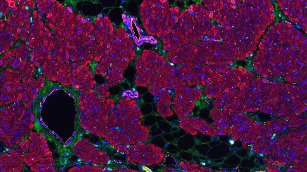
IMS image of a pediatric liver, courtesy of Dr. Hua Tian
CODEX image of the left atrium from Drs. Kai Tan and Kyung Ahn at Children’s Hospital of Philadelphia
