Image of the Week
Some of the most amazing things to come out of the HuBMAP Consortium are the images of healthy human tissues generated by our researchers.
Here, we collected them in one place to celebrate the work of these talented individuals.
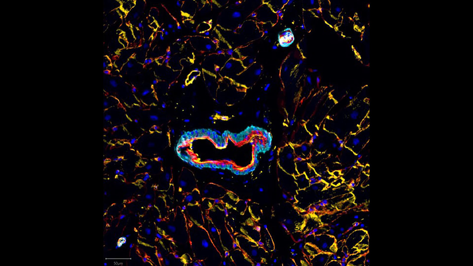
CellDIVE image of blood vessels from Drs. Liz McDonough and Fiona Ginty at GE Research
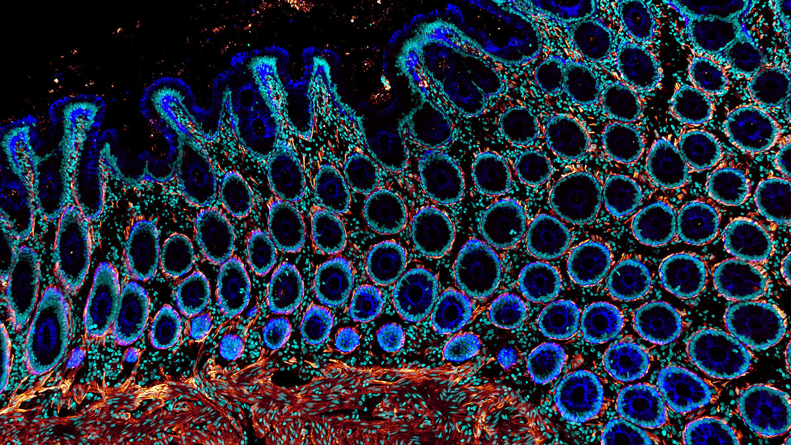
Confocal microscopy image of human colon from Dr. Andrea Radtke of the Germain lab at NIAID
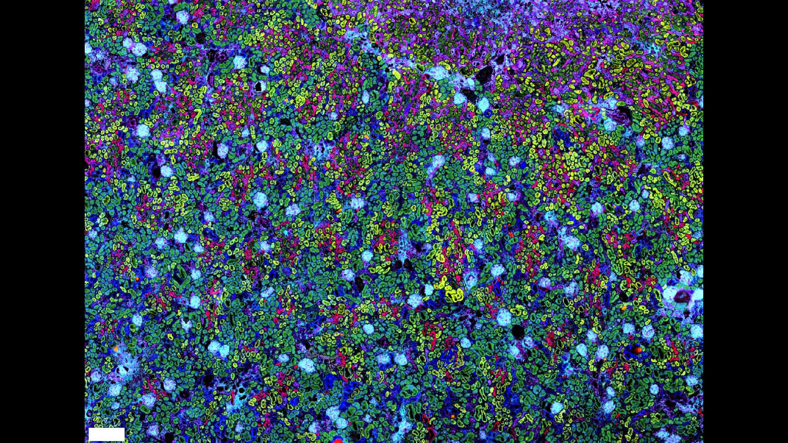
MALDI IMS image of human kidney from Dr. Martin Dufresne of Vanderbilt University
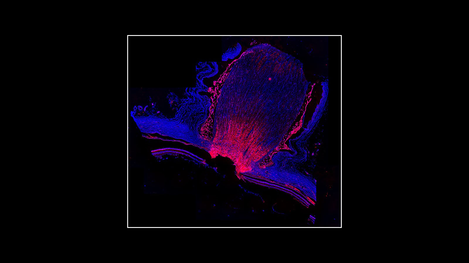
Immunofluorescence image of the human optic nerve, courtesy of Dr. Angela Kruse of Vanderbilt University
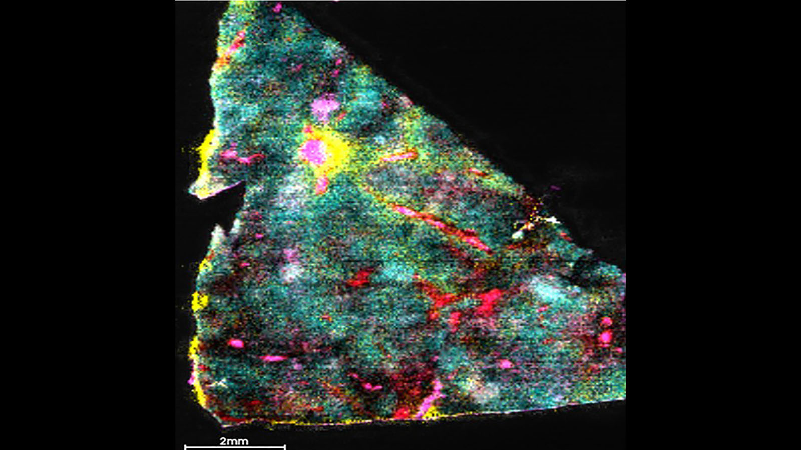
DESI image of lipids within a human liver, courtesy of Dr. Presha Rajbhandari at Columbia University
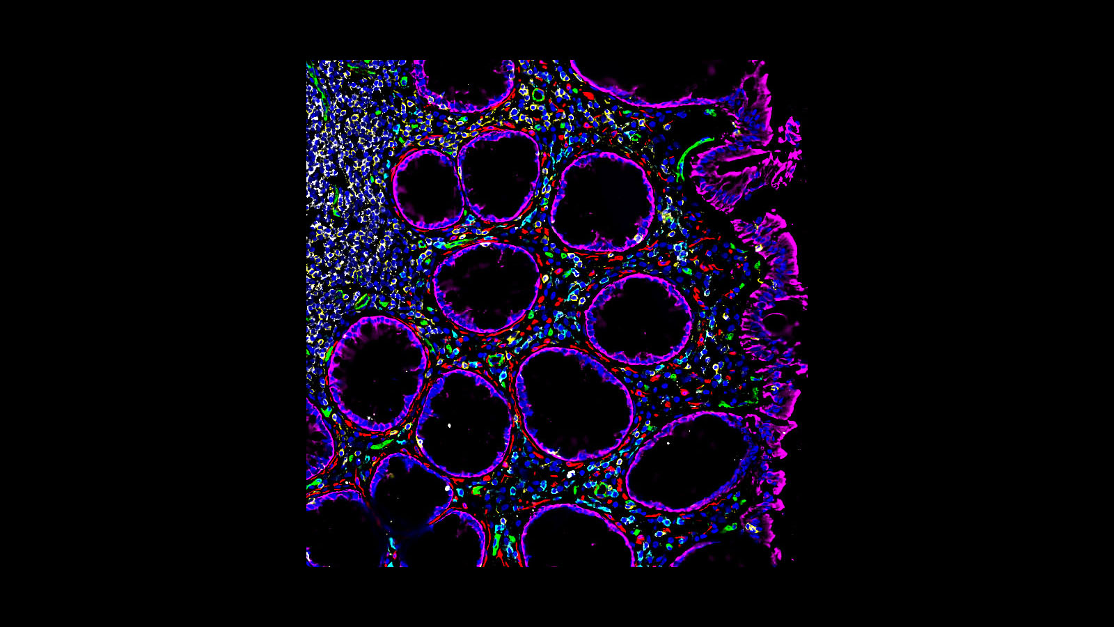
CODEX image of a healthy human colon courtesy of Dr. John Hickey of Garry Nolan's lab at Stanford
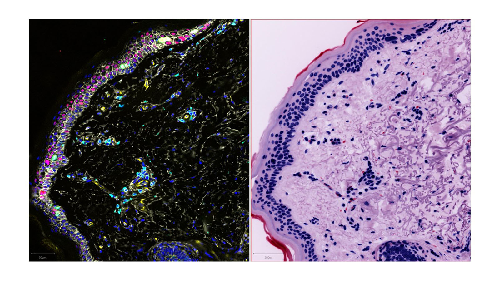
CellDIVE and H&E staining image of photoaging in response to sun exposure, courtesy of Dr. Fiona Ginty's lab at GE Research
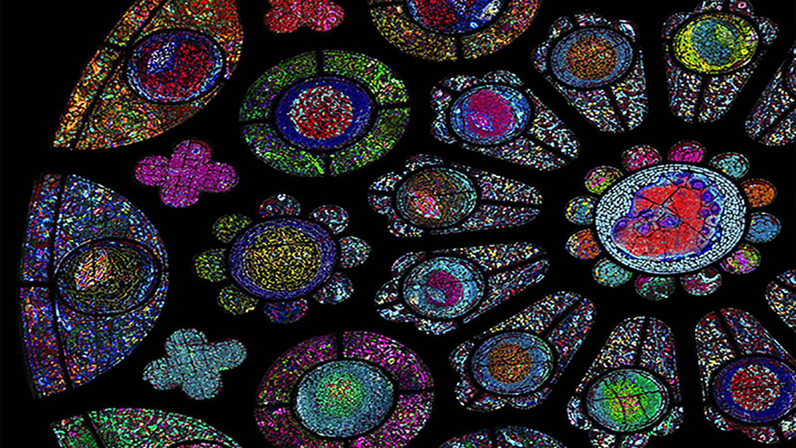
Highly multiplexed immunofluorescent image from a collaborative effort from 6 components of HuBMAP and external researchers
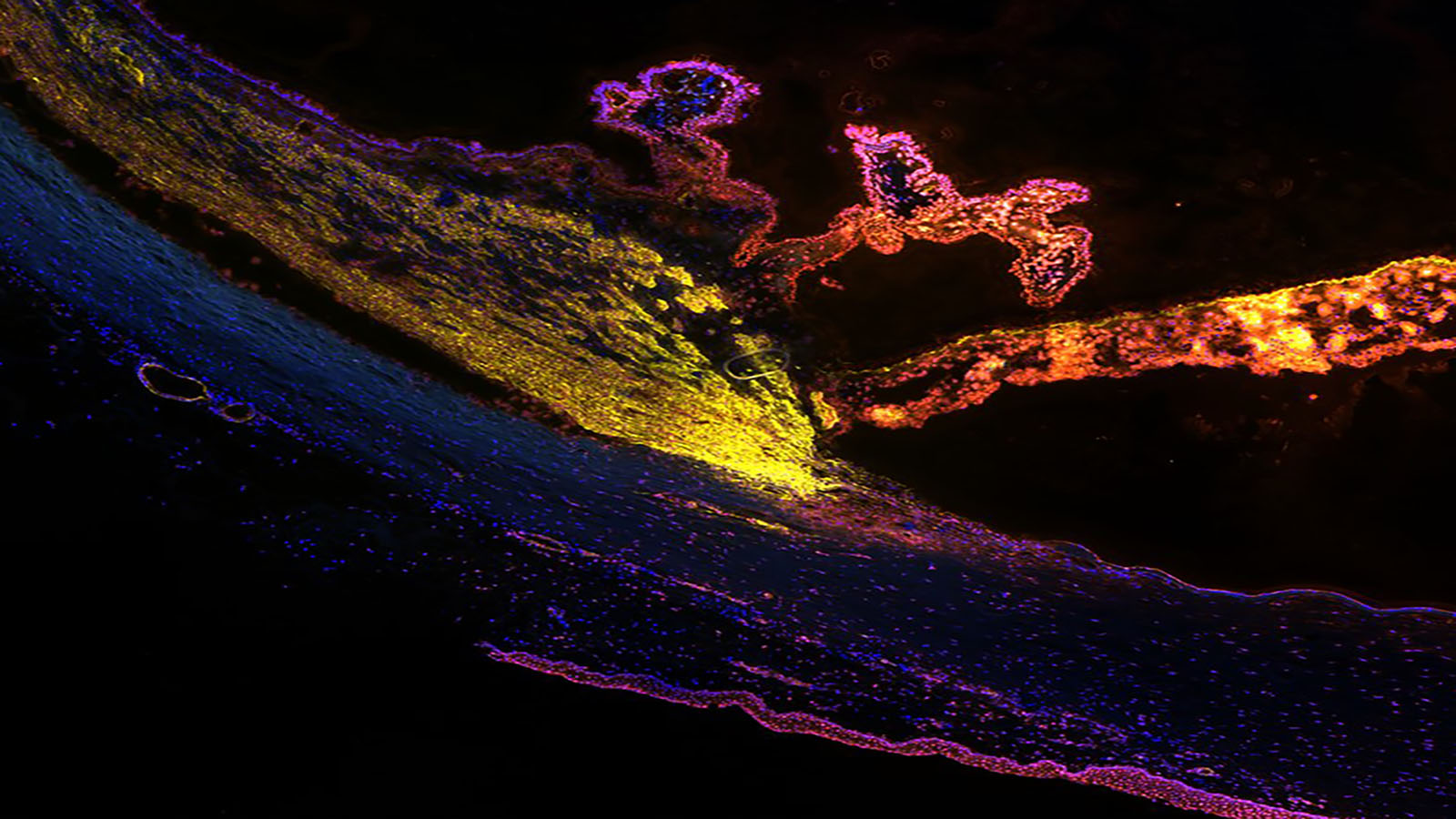
Multiplexed immunofluorescence image of the eye, courtesy of Dr. Angela Kruse of Vanderbilt University
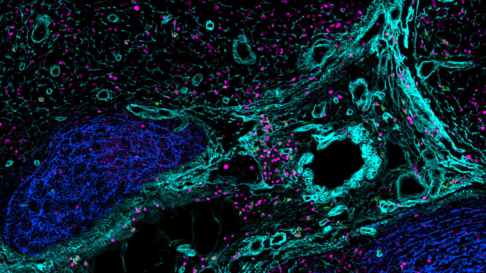
IBEX image of B cells, courtesy of Andrea Radtke at NIAID
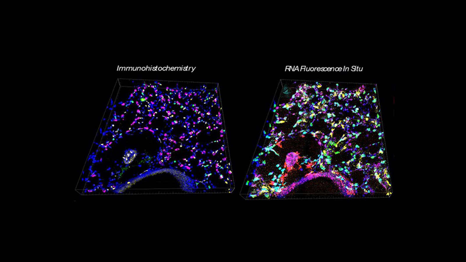
IHC and RNA fluorescence images of mouse lung, courtesy of Peter Chou at Stanford University
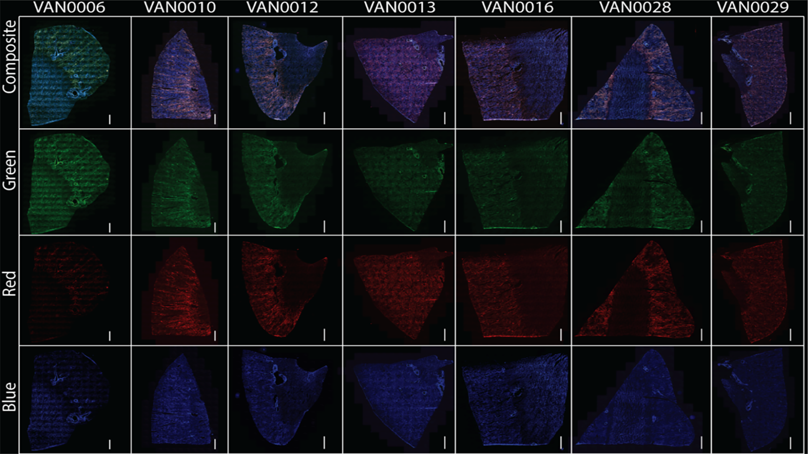
Autofluorescent images of kidneys courtesy of Dr. Elizabeth Neumann of Vanderbilt University


