
The Human BioMolecular Atlas Program (HuBMAP)
The Human BioMolecular Atlas Program (HuBMAP)
Image of the Week
Some of the most amazing things to come out of the HuBMAP Consortium are the images of healthy human tissues generated by our researchers.
Here, we collected them in one place to celebrate the work of these talented individuals.
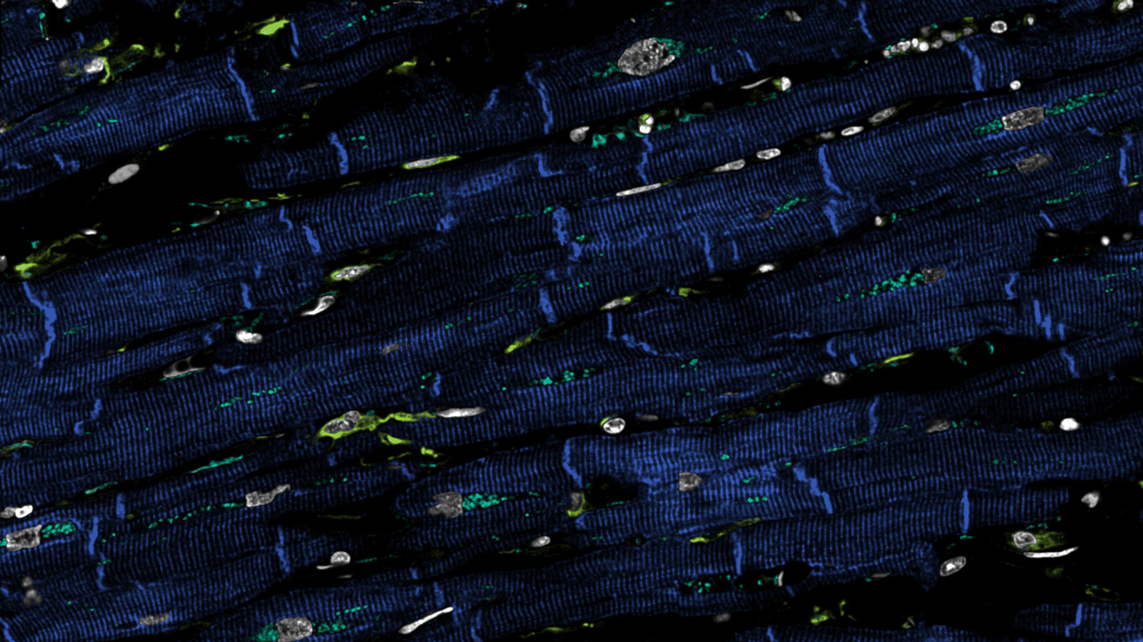
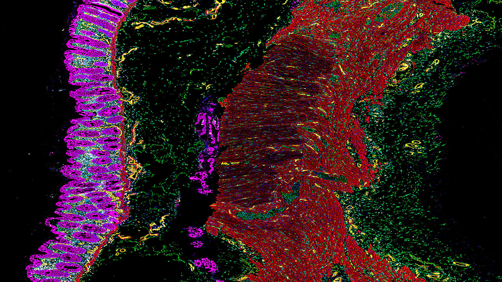
Confocal microscopy image of human heart, courtesy of Dr. Andrea Radtke of the Germain Lab at NIAID
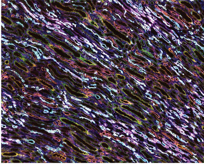
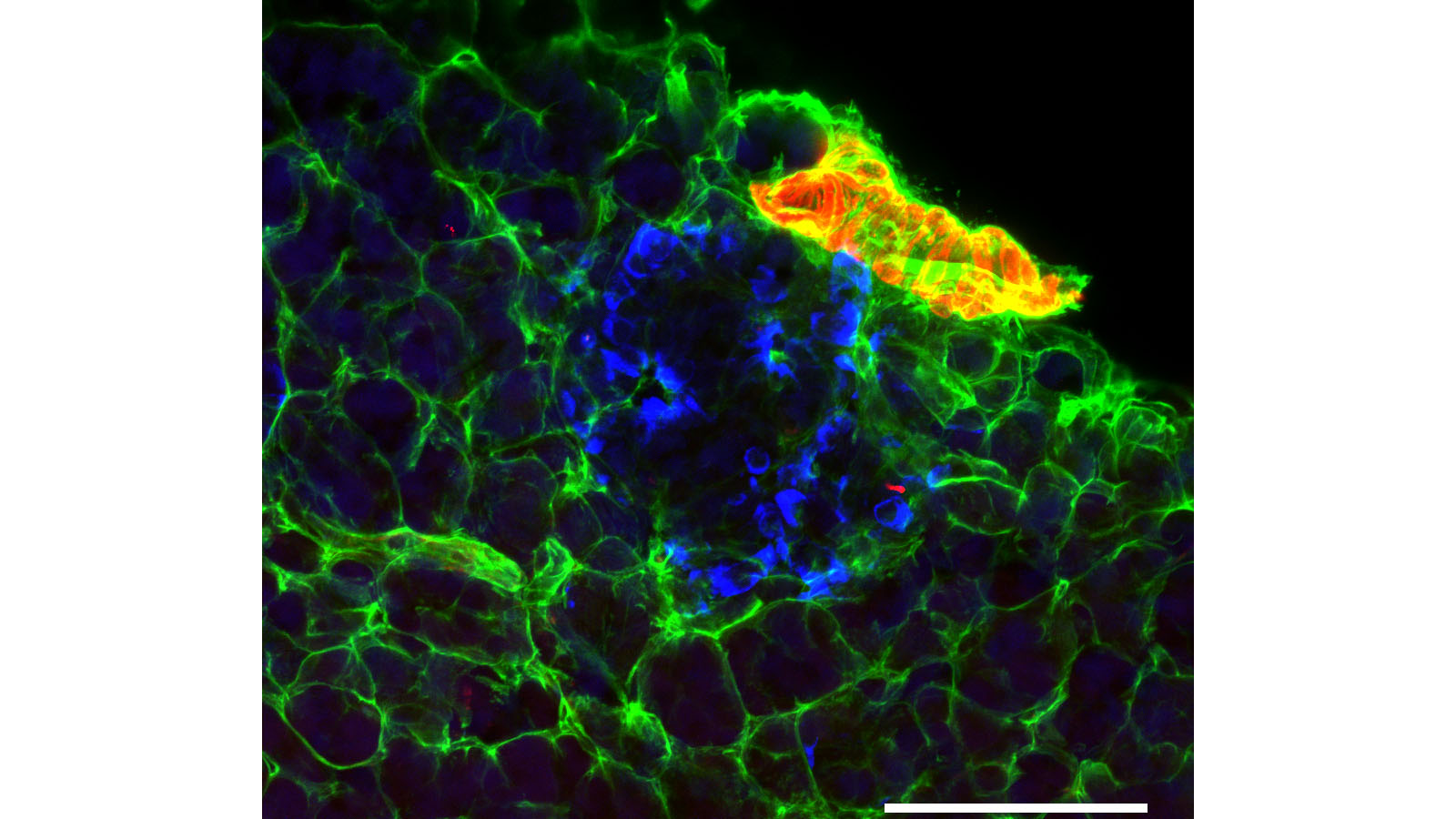
CODEX of human medullary rays in the kidney, courtesy of Dr. Elizabeth Neumann of Vanderbilt
RNAscope image of portal triad in the liver, courtesy of Aubrianna Decker at Columbia University
Mass spec image of the portal triad in human kidney, courtesy of Dr. Hua Tian from Penn State

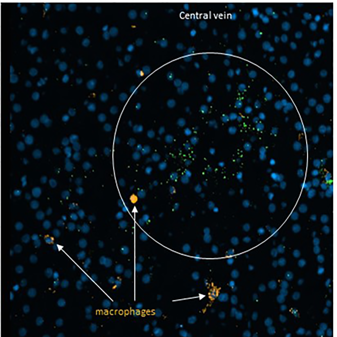
IBEX image of healthy human spleen, courtesy of Dr. Andrea Radtke of the Germain Lab at NIAID
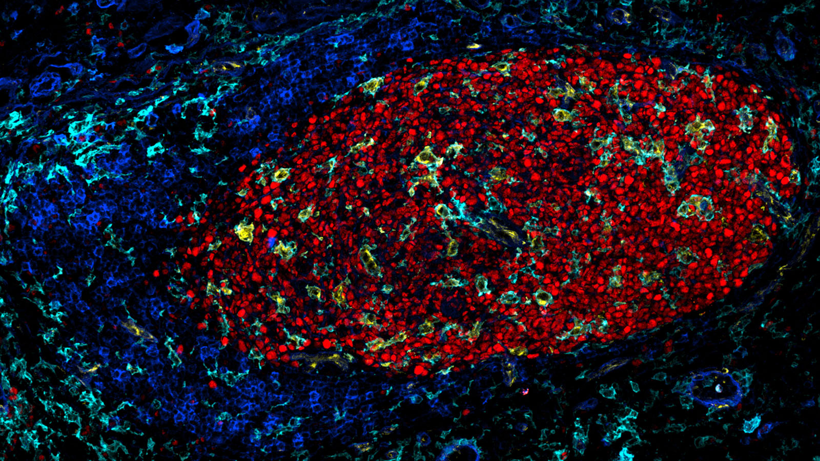
IBEX image of germinal center of human lymph nodes, courtesy of Dr. Andrea Radtke of the Germain Lab at NIAID
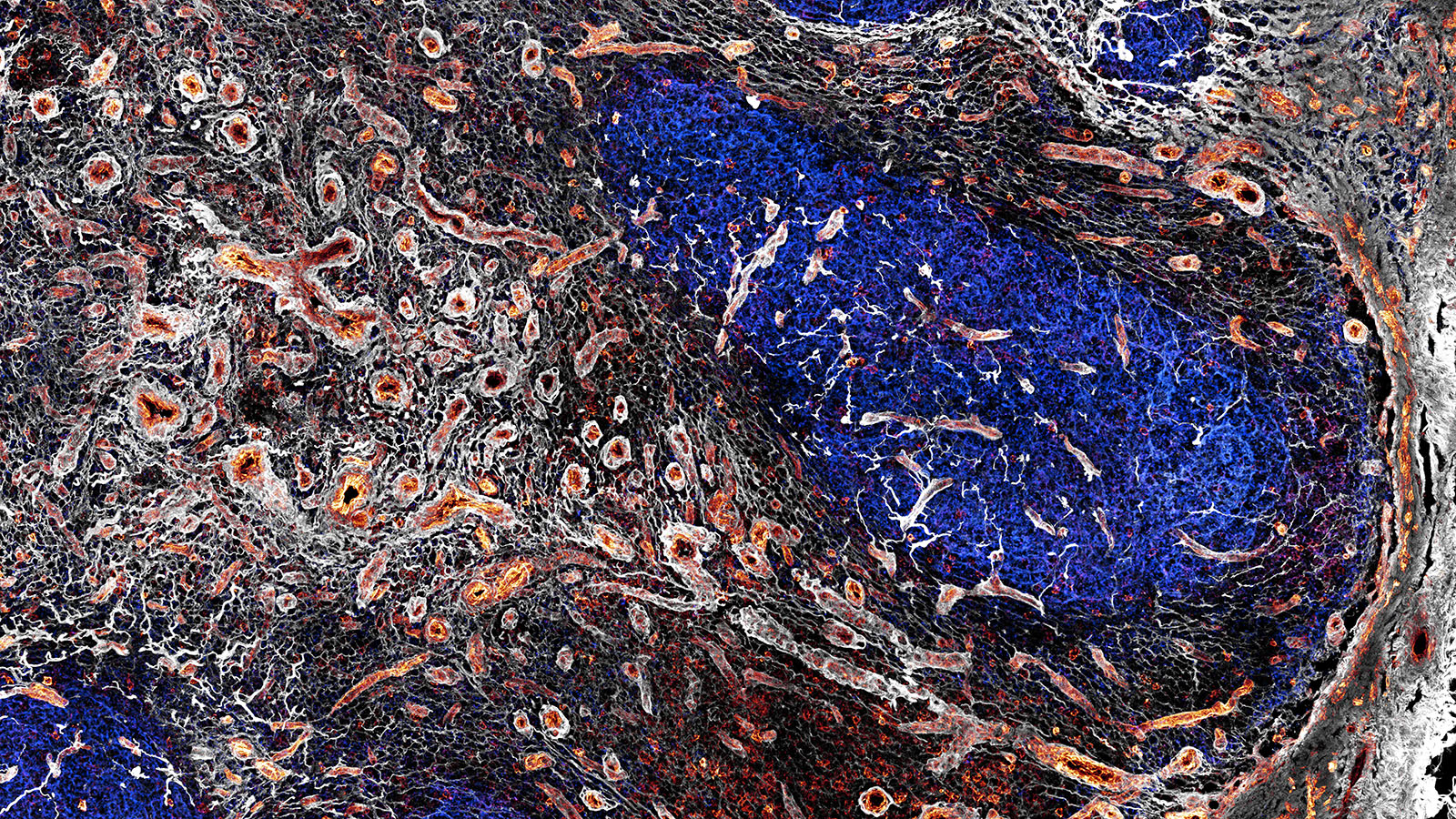
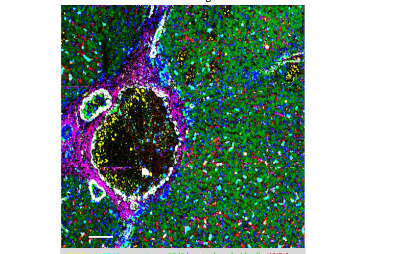
IBEX image of healthy human lymph nodes, courtesy of Dr. Andrea Radtke of the Germain Lab at NIAID
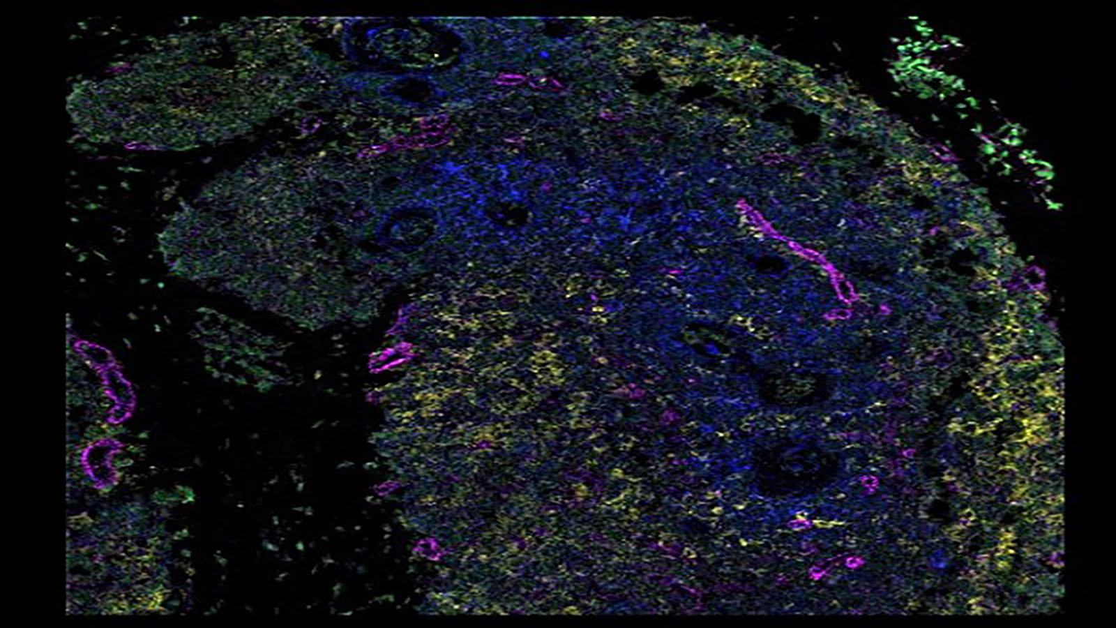
IMC image of human thymus, courtesy of Michelle Daniel of the Bodenmiller Lab
Lightsheet microscopy image of heathy human pancreas, from Katelyn Carty at the University of Florida
CODEX image of healthy human colon, from Dr. John Hickey of the Nolan Lab at Stanford
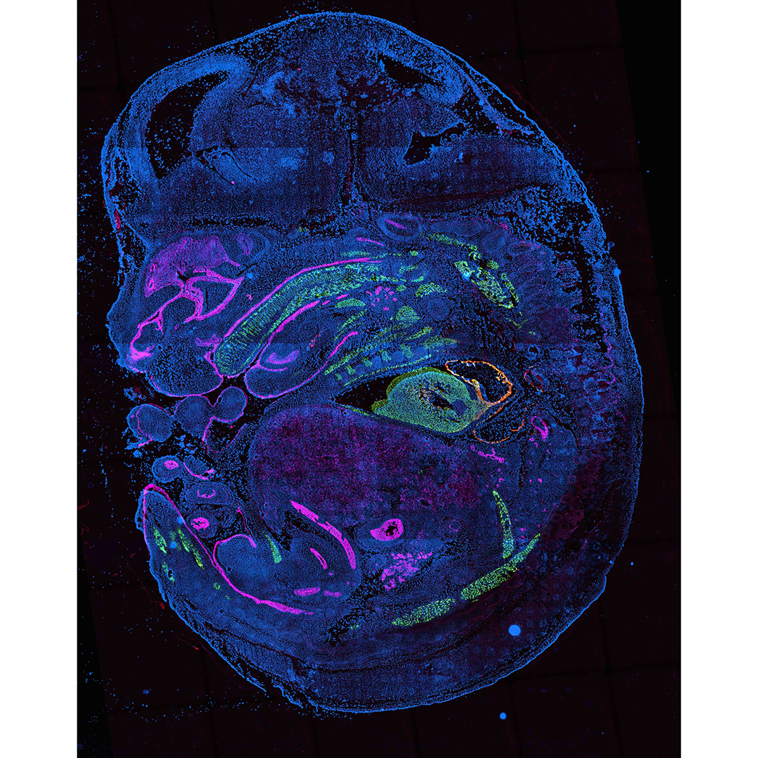
Sci-Space image of mouse embryo (stage E14), from Dr. Cole Trapnell at UW
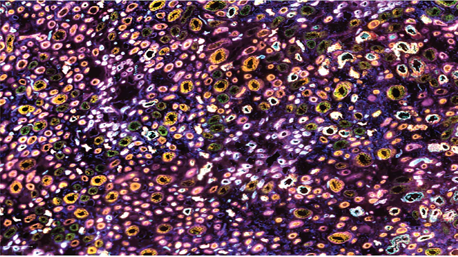
CODEX image of human kidney medulla, from Dr. Elizabeth Neumann at Vanderbilt
