
The Human BioMolecular Atlas Program (HuBMAP)
The Human BioMolecular Atlas Program (HuBMAP)
Image of the Week
Some of the most amazing things to come out of the HuBMAP Consortium are the images of healthy human tissues generated by our researchers.
Here, we collected them in one place to celebrate the work of these talented individuals.
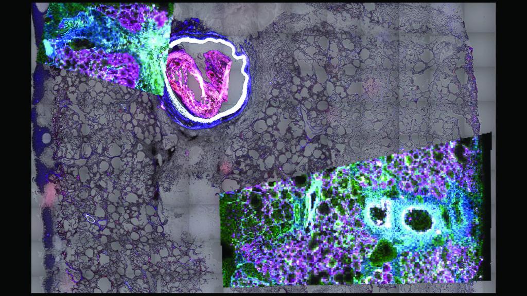
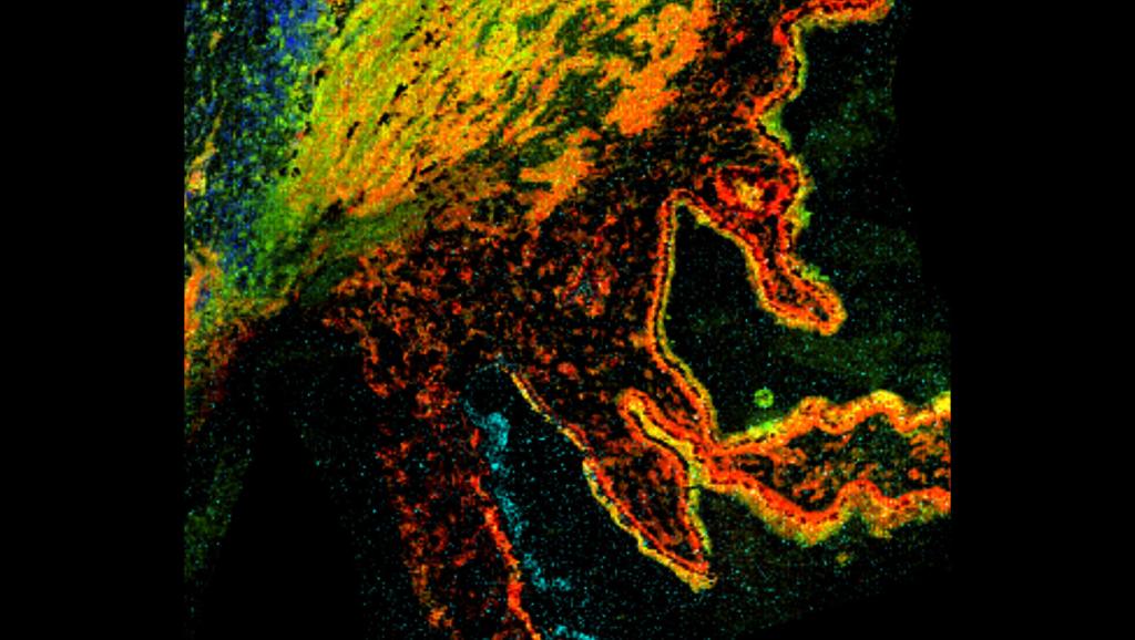
This is a MALDI-MSI & autofluorescence image of the lipids inside the central portion of the left upper lobe of the lung, showing the large bronchus and smaller bronchioles by HuBMAP researcher Dr. Brittney Gorman at Pacific Northwest National Laboratory
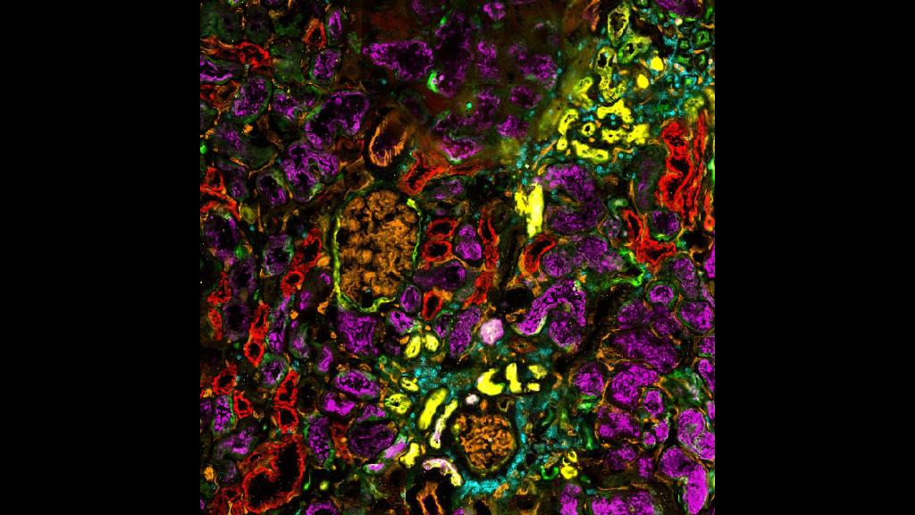
CODEX image of a human kidney from Drs. Tarek Ashkar, Angela Sabo, and Daria Barwinska at Indiana University
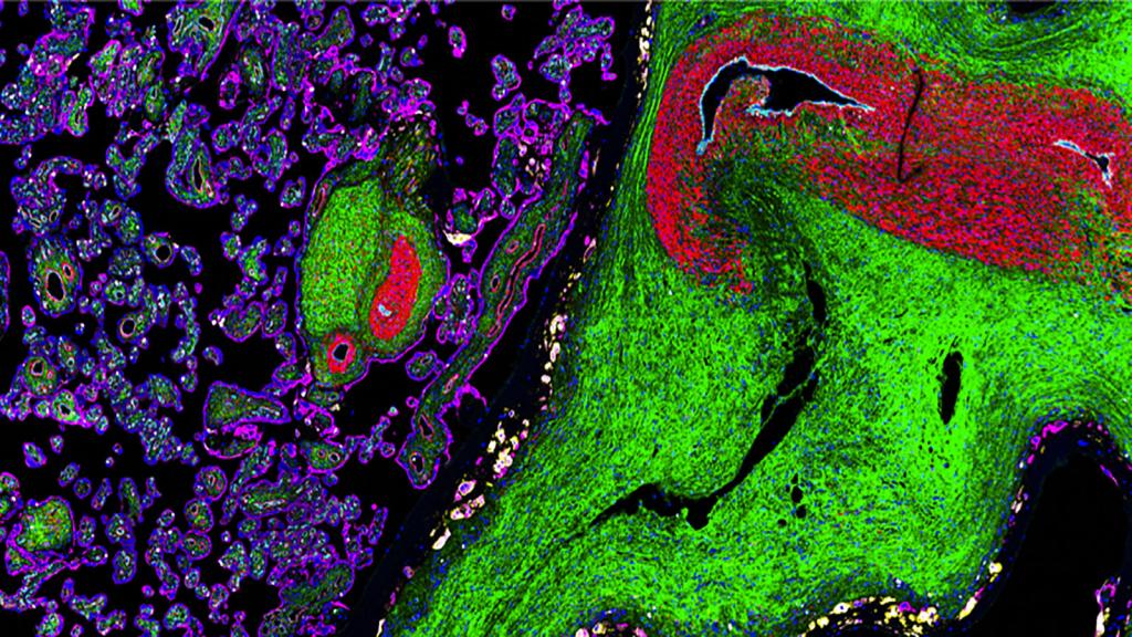
Organ Mapping Antibody Panel (OMAP) of the placenta from HuBMAP researchers Drs. Santhosh Sivajothi and Ramalakshmi Ramasamy at The Jackson Laboratory
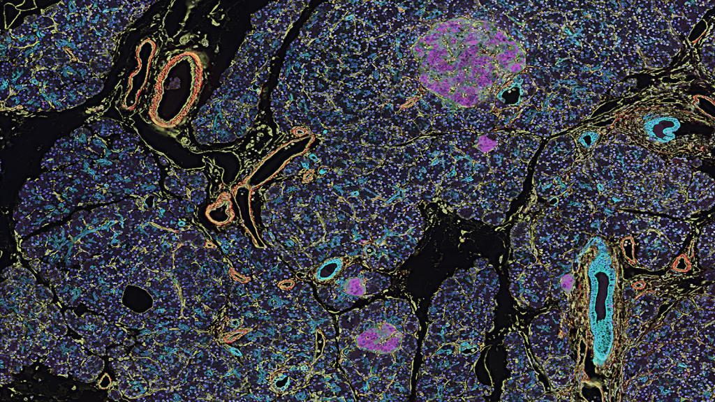
CODEX image of a healthy pancreas from Drs. Frida Björklund & Anna Martinez Casals at Emma Lundberg’s lab at Stanford
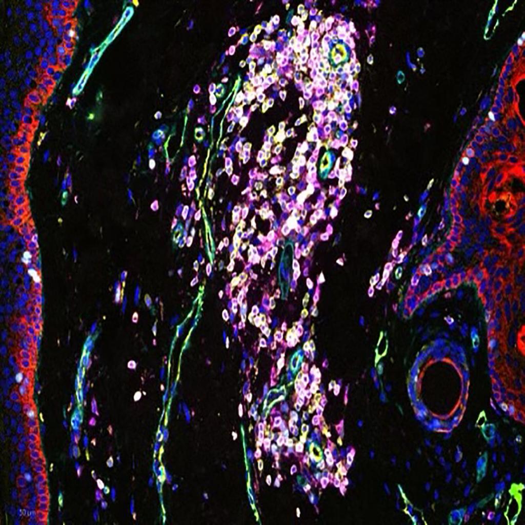
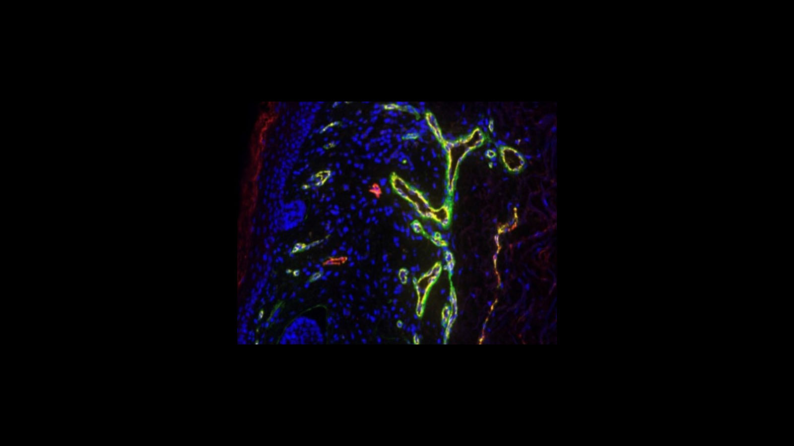
CellDIVE image of the skin, courtesy of Dr. Liz McDonough at GE
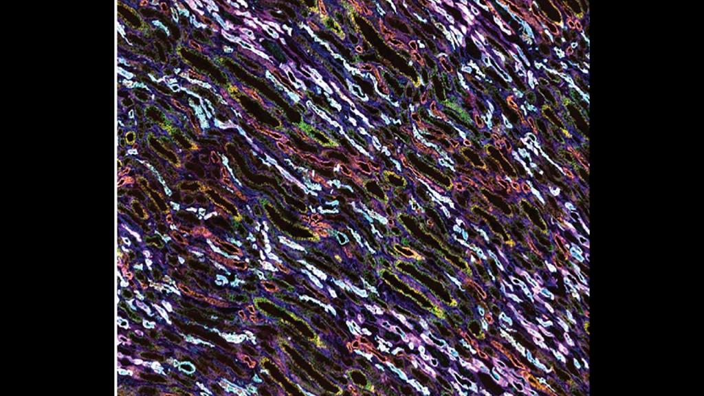
This is a CODEX MxIF image of medullary rays inside the kidney made by HuBMAP researcher Dr. Elizabeth Neumann at Vanderbilt TMC
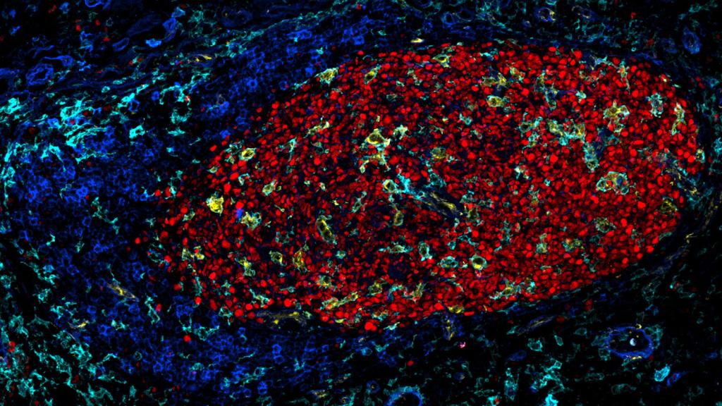
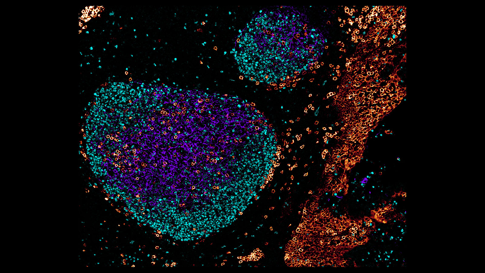
IBEX image of a germinal center of a lymph node by Dr. Andrea Radtke at National Institute of Allergy and Infectious Diseases’ Germain lab.
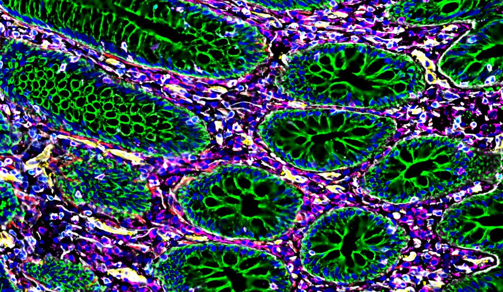
Organ Mapping Antibody Panel (OMAP) of the intestine by Dr. John Hickey at Stanford University
MALDI image of a ciliary process in a human eye from Dr. David Anderson at Vanderbilt TMC
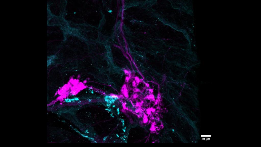
3D microscopy image of a neural node within the pancreas by Dr. Angela Kruse at Vanderbilt University
Confocal microscopy image of a human tonsil by Dr. Andrea Radtke in the Germain lab at NIAID
Cell DIVE multiplexed immunofluorescent image of skin courtesy of Dr. Fiona Ginty, Chrystal Chadwick, and Liz McDonough at GE Research
