
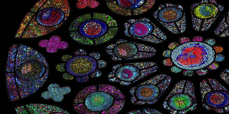
The human body is made up of trillions of cells, each having a specialized role, and each affected by its environment and neighboring cells. The NIH Common Fund’s Human BioMolecular Atlas Program (HuBMAP) is a consortium of over 300 researchers studying the spatial organization of cells within tissues to better understand normal function of organs in health and disease. As part of that effort, HuBMAP is developing and optimizing new technologies to study cells within their native states in different tissues, such as “multiplex antibody-based imaging.” In methods like this one, antibodies are used for their ability to bind to proteins that only appear in a certain kind of cell. Attached to one end of the antibody is a molecule that serves as a marker, making the location of the antibody visible to imaging equipment like high-tech microscopes. Scientists can look for these markers and identify the types of cells in a tissue sample based on the proteins recognized by the antibodies. Many different antibodies are used to recognize different kinds of proteins, which is called a multiplex approach. Recently, different components of HuBMAP came together to write a primer, or instruction manual, to describe the resources, considerations for obtaining high-quality imaging data, and expert knowledge needed to assist other researchers in choosing the best multiplexed antibody-based imaging method to map proteins in tissues. The primer includes advice on choosing antibodies and experimental workflows, as well as suggestions for large dataset management for these new methods. Additionally, the authors highlighted the need to integrate multiplexed imaging data with other biomolecule data types for more robust analyses. Such analyses that combine different types of data to form a more complete picture, will allow researchers to measure many different biomolecule types, like RNA and protein, within the same sample. This will give a better understanding of internal cellular functions, as well as the influence of neighboring cells on a particular cell in a certain location.
The primer, which appears as a perspective paper in the journal Nature Methods, outlines the challenges and advancements involved in multiplexed antibody imaging. It is hoped its guidance and other advances in HuBMAP will contribute to the development of tissue maps that aid in understanding normal tissue and organ functioning, disease progression, and response to treatment in different tissues.
Spatial Mapping of Protein Composition and Tissue Organization: A Primer for Multiplexed Antibody-Based Imaging. John W Hickey, Elizabeth K Neumann, Andrea J Radtke, Jeannie M Camarillo, Rebecca T Beuschel, Alexandre Albanese, Elizabeth McDonough, Julia Hatler, Anne E Wiblin, Jeremy Fisher, Josh Croteau, Eliza C Small, Anup Sood, Richard M Caprioli, R Michael Angelo, Garry P Nolan, Kwanghun Chung, Stephen M Hewitt, Ronald N Germain, Jeffrey M Spraggins, Emma Lundberg, Michael P Snyder, Neil L Kelleher, Sinem K Saka. Nat Methods. 2022 Mar;19(3):284-295. doi: 10.1038/s41592-021-01316-y. Epub 2021 Nov 22. PMID: 34811556 DOI: 10.1038/s41592-021-01316-y
This work is supported by U54 HG010426, U54 DK120058, UH3 CA246635, UH3 CA246594, UH3 CA246633, NIAID (Andrea Radtke and Ron Germain), NCI (Stephen Hewitt)
