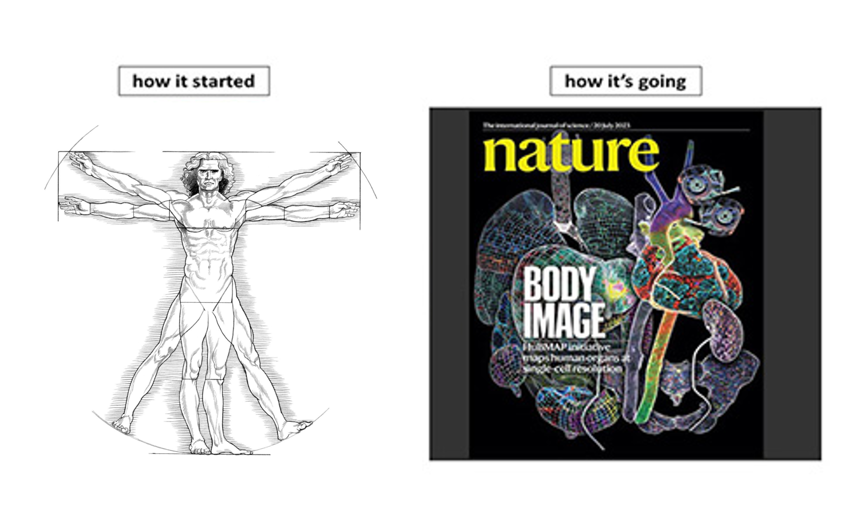
Since humans first made the connection between the ailments they were feeling and issues with their bodies, they have been fascinated by the structure and function of the body, and how it relates to human health. Over time, people discovered that cells make up living organisms, and those cells have specific functions performed in that organism to keep them healthy. Those cells can be influenced by the neighboring cells and the environment around them. If something causes a disruption within or near cells, it might lead to damage or a disease. In order to study cells and their behavior within an organ or tissue, the NIH Common Fund’s Human BioMolecular Atlas Program (HuBMAP) established a consortium of over 400 researchers from 60 institutions using cutting-edge techniques to study the spatial organization of cells within tissues to better understand normal function of organs in health and disease. The most recent findings of the HuBMAP Consortium researchers have been brought together in a collection of 13 articles published by Nature family journals, which you can check out here (Nature Landing Page). HuBMAP researchers have brought together data on RNA, proteins, and metabolites in human organs at single-cell resolution to generate an open-access data platform for researchers to study the inner workings of the cells and how they affect health.
Between the renaissance in technology necessary for this work to happen, and the beautiful images that it has generated, even Leonardo Da Vinci himself would give a Mona Lisa smile for HuBMAP’s body of work!


