
Extracellular vesicles are particles that exist outside of cells and are present in different bodily fluids. Small extracellular vesicles have been found in all samples of body fluids, making them a great candidate for a biomarker, which is substance whose presence can indicate disease or environmental exposure. Small extracellular vesicles can serve as biomarkers for diseases like cancer, infections, and neurological diseases. The ability to capture extracellular vesicles in lab settings an important foundation to studying their nature and behavior in the human body.
Current methods of extracting extracellular vesicles are difficult to conduct in lab settings. Researchers in the NIH Common Fund Extracellular RNA Communication Consortium (ERCC) have developed a separation method to recover more small extracellular vesicles from a sample than the current gold standard method, which could optimize important biomedical research.
The gold standard method to isolate small extracellular vesicles includes centrifugation, a process by which a liquid is rapidly spun to separate its components based on size. However, centrifugation involves long processing times, low yield of small extracellular vesicles, high equipment costs, and the potential of incomplete separation of the components of the sample.
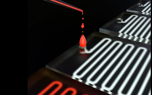
The new isolation process developed by ERCC researchers is conducted on a microscopic device. The microscopic device has different sized channels which separate fluid components by size. For example, the device can separate blood into white blood cells, red blood cells, and platelets. Then, using a microscopically thin filter, the small extracellular vesicles can be separated from the different components. This process takes less time and is more precise than previous methods. Compared to the gold standard centrifugation method, the microfluidic device yielded 30 times more small extracellular vesicles than ultracentrifugation.
This new method provides opportunities for new therapies and enhanced disease research. When testing this new method in clinical practice, higher concentrations of small extracellular vesicles were found in 20 cancer patients compared to 20 healthy donors, indicating the importance of future applications for cancer research and clinical research overall. Accurately capturing small extracellular vesicles is crucial to studying their patterns in disease detection.
Reference: Yingchao Meng, Yanan Zhang, Marcel Bühler, Shuchen Wang, Mohammad Asghari, Alessandra Stürchler, Bogdan Mateescu, Tobias Weiss, Stavros Stavrakis, and Andrew J. Demello. Direct isolation of small extracellular vesicles from human blood using viscoelastic microfluidics.Sci. Adv.9,eadi5296(2023).DOI:10.1126/sciadv.adi5296
