

Glycoscience
Glycoscience
Science Highlights Archives
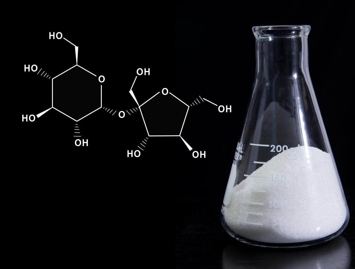
New Method for Generating Carbohydrates Sweetens the Deal
 Common Fund Glycoscience grantee Dr. Eric Jacobsen and collaborators at Harvard University have developed a new approach to allow more scientists to study important biological sugar molecules, advancing research and accelerating progress in understanding the role of sugars in human health and disease. Carbohydrates, or sugars, are naturally occurring chemical compounds present throughout every living cell. Although carbohydrates play key roles in several biological processes, including photosynthesis and metabolism, they have remained largely understudied because generating carbohydrates in a laboratory is challenging. Carbohydrates are generated through chemical synthesis, the construction of complex chemical compounds from simpler ones. Due to their complicated three dimensional structure, or stereochemistry, carbohydrates are especially complex compounds, which makes their chemical synthesis difficult and erratic.
To overcome this challenge, Dr. Jacobsen and collaborators developed a method to generate complex carbohydrates in a stereospecific manner using a unique chemical catalyst. A catalyst is a substance that enables a chemical reaction to proceed at a faster rate or under different conditions than otherwise possible. For example, a glycosylation enzyme is a biological catalyst that attaches carbohydrates to other molecules in living cells. Interestingly, the chemical catalyst developed by Dr. Jacobsen’s group mimics biological glycosylation enzymes. Using a chemical catalyst instead of enzymatic catalyst can be useful when large amounts of carbohydrate are required for research. This is because biological enzyme catalysts may be unstable or hard to isolate. Additionally, although there are existing enzymes and methods to synthesize carbohydrates, these approaches are not predictable or straight forward. This means that every time a scientist wants to synthesize a carbohydrate to study it, the expertise of a carbohydrate synthesis specialist is required. This new approach has the potential to enable the study of glycoscience by non-specialists, a major goal of the Common Fund’s Glycoscience program and an important step in advancing the field of glycoscience.
In the News: Sweeter hookups between sugars using a macrocyclic catalyst (link is external)
Reference: Macrocyclic bis-thioureas catalyze stereospecific glycosylation reactions. Park Y, Harper KC, Kuhl N, Kwan EE, Liu RY, Jacobsen EN. Science. 2017 January 13.
Exciting New Method for Probing Glycans in Clinical Specimens!
 Glycans, or sugars, are attached to many cellular proteins and play important roles in a variety of cellular processes. All cells carry an array of glycans that modulate molecules that are critical to the development and function of multicellular organisms. Additionally, the glycans present on cellular proteins change with disease state, making glycans useful as disease biomarkers (a measurable substance in an organism whose presence is indicative of disease, infection, or environmental exposure). However, analysis of glycans in clinical specimens is limited because current methods tend to require higher concentrations of glycan than is typically available from a clinical specimen. To overcome this challenge, Common Fund Glycoscience Program grantee Dr. Brian Haab and collaborators have developed a new method that allows for the detection of small sample of human plasma (the clear, liquid part of the blood, composed mainly of water and proteins, in which the blood cells are suspended), as well as precision comparisons over multiple samples. Using this new method, called on-chip glycan modification and probing (on-chip GMAP), proteins with glycan modifications were immobilized on a surface and the amount of glycans on the proteins was detected. They precisely analyzed glycans found on proteins, as well as compared the presence and structure of glycans over multiple samples and disease states. As a proof of principle, on-chip GMAP was used to detect the cancer biomarker MUC5AC, a glycoprotein extracted from human plasma. The amount of protein required was 25 – 50 times lower than previous methods. This new method can be used as a platform for analyzing small amounts of a given glycoprotein in clinical specimens and determining how these glycoproteins change in disease.
Reference: Characterizing Protein Glycosylation through On-Chip Glycan Modification and Probing. Reatini BS, Ensink E, Liau B, Sinha JY, Powers TW, Partyka K, Bern M, Brand RE, Rudd PM, Kletter D, Drake RR, Haab BB. Analytical Chemistry. 2016 Nov 3
Novel Tool for Quantitative Detection of O-GlcNAc Modifications on Specific Proteins
 Proteins are large biomolecules that are required for the structure and function of the body’s cells and tissues. The human body contains somewhere between 17 to 18 thousand different proteins. The function of any one of these proteins can be changed by the addition or removal of chemical modifications to the protein. These modifications can exert profound effects on a proteins activity, cellular localization, and/or its interaction with other proteins and biomolecules. O-GlcNAcylation is a protein modification that has been implicated in multiple disease process including cancer, diabetes, and Alzheimer’s disease. However, O-GlcNAcylation of proteins can only be currently analyzed using complicated procedures that are accessible to just a few scientists who have highly specialized and expensive equipment. To overcome these hurdles, Common Fund Glycoscience grantee Dr. Carolyn Bertozzi has developed a new method, termed Glyco-seek. Taking advantage of a common laboratory method (polymerase chain reaction, PCR), Glyco-seek is more convenient than existing methods for the detection of O-GlcNAcylated proteins. In addition to being more amenable to most research laboratories, Glyco-seek also shows ultrahigh sensitivity when compared to current techniques. This method has potentially broad applications for the study of other forms of glycosylation, as well as the potential to enable the broader scientific community to easily analyze glycosylated proteins.
Reference: Glyco-seek: Ultrasensitive Detection of Protein-Specific Glycosylation by Proximity Ligation PCR. Journal of the American Chemical Society. 2016 July 25.
New Low Cost, Adaptable Method for Purifying Carbohydrates
 Given the complexity of carbohydrates, their use in biological studies requires that they be of the highest level of purity to ensure rigorous and reproducible results in biological experiments. Inadequate access to pure carbohydrates has long hindered the study of their biology and impact on human health, as well as their therapeutic value. To address roadblocks in carbohydrate purification, Glycoscience grantees Drs. Nicola Pohl and Milos Novotny, from Indiana University, have developed a new protocol for the purification of carbohydrates. This low cost methodology addresses biologists concerns for access to glycans of greater than 99% purity.
Reference: Protocol for the purification of protected carbohydrates: toward coupling automated synthesis to alternate-pump recycling high-performance liquid chromatography.Chemical Communications. 2016 October 24.
New Strategies for the Facile Production of Natural Glycans to Accelerate their Study and Use
 Glycomics is the study of all glycan structures of a given cell type or organism. Rigorous examination of any given glycome requires the efficient release, recovery, and analysis of all glycans from that cell type or organism. Subsequent study of individual glycans further requires they be available in sufficient quantity and that methods and standards for their identification be available. The chemical and enzymatic synthesis of thousands of naturally occurring glycans is a significant challenge in the field of glycomics. Toward overcoming this challenge, Glycoscience Program awardees Drs. Xuezheng Song and Richard Cummings have developed a new facile strategy for obtaining naturally occurring glycans from biological tissues, including mouse and pig. Traditional methods for preparing natural glycans are both lengthy and expensive. Further, they rely on enzymatic digestions that typically produce limited quantities of glycans (milligrams). To overcome these obstacles, Drs Song and Cummings have developed a new strategy, called oxidative release of natural glycans (ORNG), which quickly releases glycans from biological samples (both proteins and lipids) in kilograms quantities, alleviating the need for expensive enzymes and prolonged digestion times. This new and innovative approach is a significant step forward because it uses an inexpensive and mild reagent (household bleach) to “substantially improve the simplicity and accessibility” of mammalian glycomes, allows further study of their functions, and provides them in significant quantity for development of tools.
Reference: Oxidative release of natural glycans for functional glycomics. Song X, Ju H, Lasanajak Y, Kudelka MR, Smith DF, Cummings RD. Nature Methods 2016 June 13.
Studying Sugars on a Chip
 Proteins are key biological components of almost every action in our cells. They come in all different shapes and sizes and often work together as the machinery our cells use to get jobs done. An important part of biology is how glycans (sugars) in our body interact with proteins to change, enhance, or stop how proteins work. However, it is difficult to uncover information about how glycans interact with proteins due to the complexity of glycan structure and the large amount of biological material needed to study the protein/glycan interaction.
The goal of the Common Fund Glycoscience program is to make glycan biology more accessible to the biomedical research community. A paper from the lab of Glycoscience researcher Brian Haab describes a new method that combines typical experimental approaches using glycan-bound proteins purified from cells and computational analysis to gain information on the complex structures of the glycans using only a small amount of material. The researchers placed droplets of pure proteins on a microchip where enzymes chewed apart the complex glycans attached to the proteins into smaller pieces that were detected using another technique. Computer algorithms were then used virtually to put the pieces of the glycan puzzle back together to see what it looked like when it was attached to the protein. Only needing a small amount of material makes this technique more accessible because it could be applied to precious samples from human patients or used by labs that are not equipped to generate large quantities of glycan-bound proteins for study.
Reference:
Deciphering Protein Glycosylation by Computational Integration of On-chip Profiling, Glycan-array Data, and Mass Spectrometry. Klamer Z, Hsueh P, Ayala-Talvera D, and Haab B. 2019, Molecular and Cellular Proteomics (18) 28-40 https://doi.org/10.1074/mcp.RA118.000906 (link is external)
Researchers Develop New Automated Method to Build Complex Sugars
 The study of glycans (sugars) within our body and how they interact with different biological molecules, like proteins, provides important information on many of our bodies’ activities, such as fighting off infections or cell-to-cell communication. Despite the value in studying glycans, glycobiology is not a field many researchers pursue as it is very difficult to create glycans artificially or to monitor natural glycans in living cells. A goal of the Common Fund Glycoscience program is to enable any investigator to study how glycans impact their area of study, even if he or she is not a glycobiology expert.
An important step in making glycans more accessible to the broader research community is to create easier ways to generate them in a laboratory. Glycans come in a variety of forms in biology, ranging from simple to complex structures that have many sugars put together in intricate branching networks. A new way to create complex sugar structures was developed by Glycoscience awardee Dr. Geert-Jan Boons and colleagues that allows for automated development of complex glycans – even human milk glycans – using enzymes found in biology. A paper describing the method was published in Nature Chemistry. In the paper, the scientists describe how to efficiently purify newly synthesized glycans, greatly reducing the time and improving the yield. This new method will grant easy access to different classes of complex glycans, allowing both experts and non-glycobiology researchers to study how different glycan forms impact a wide range of biology, such as neurodevelopment, diseases of the nervous system, and cancer metastasis.
Reference:
An automated platform for the enzyme-mediated assembly of complex oligosaccharides. Li T, Liu L, Wei N, Yang JY, Chapla DG, Moremen KW, and Boons GJ. 2019, Nature Chemistry, (3) 229-236
Glycoscience experts want you to study glycans!
 Glycans (sugars) play an integral role in all components of human biology. They help our immune system recognize foreign invaders, allow our cells to adhere to different surfaces, and even help form the protective mucous in our nose. Despite the importance of glycans to biology, their complicated chemistry and nearly endless variety has limited the number of scientists who investigate how glycan biology affects health and disease. The overall goal of the Common Fund’s Glycoscience program is to develop new tools, resources, and information for researchers unfamiliar with glycans to incorporate them into their studies. The Glycoscience program is interested in setting up new collaborations, such as the one between Glycoscience awardee Dr. Catherine Grimes and researcher Dr. Nina Salama that allowed Dr. Salama to introduce new glycoscience experiments into her work on Helicobacter pylori, the bacterium that’s the main cause of stomach ulcers.
Glycoscience program researchers set up an “Emerging technologies in glycoscience” ask the expert workshop at the American Society for Biochemistry and Molecular Biology (ASBMB) annual meeting in April 2019 to develop more collaborations and to introduce more scientists to glycoscience. During this workshop, attendees interacted with Common Fund-funded Glycoscience experts, asking questions and learning about different tools, resources, and experimental protocols to aid them in conducting glycoscience experiments. This included techniques for identifying especially difficult glycans, probes to study glycans in bacterial cell walls, standards for glycan chemical synthesis, methods for high-throughput glycan studies, and glycoscience educational materials. The workshop was so effective, the Glycoscience program has already been invited for a repeat performance at ASBMB next year. If you missed the workshop, but are interested in the Glycoscience program, visit commonfund.nih.gov/glycoscience for program resources and announcements of future workshops and presentations.
Synthesizing sugars is no piece of cake?

Glycans are sugars that have many important biological and medical applications, such as use in clinical diagnostics and in glycan-based vaccines. However, it is difficult to obtain large enough quantities of specific glycans from nature and current methods of synthesizing glycans in the lab are time consuming and labor intensive. Current synthesis methods require skilled researchers in highly specialized laboratories and can involve as many as 100 steps. Even skilled researchers can have a very hard time making enough glycans for biomedical studies. The Glycoscience Program is developing new tools, techniques, and methods to create large amounts of glycans by simpler methods, which will make the study of glycans more accessible to the biomedical research community.
Glycoscience Program-funded investigator Dr. Geert Jan-Boons and his research team have developed a new “stop and go” approach to glycan synthesis that involves 10 or fewer steps and is designed to mimic natural glycan synthesis in the body. A process of modifying proteins by adding glycans, called “N-glycosylation,” is one of the most diverse and complex types of protein modifications. N-glycosylation is involved in many biological processes, such as neuron development, fertilization, immune regulation, and in many disease processes. This new “stop and go” strategy allows for synthesis of useful amounts of a variety of complex N-glycans without the need for special synthesis skills, providing glycans for biomedical research.
Another investigator with the Glycoscience program, Dr. Peng George Wang and his research team, developed a fully automated system to efficiently synthesize glycans using enzymatic reactions, a temperature dependent polymer, and repurposing a commercially available peptide synthesizer. Several important glycans, such as blood group antigens (i.e. the sugars on the surface of blood cells that designate A, B, and O blood types), were synthesized using this system. These experiments illustrate the important ways in which the Glycoscience Program is simplifying glycan synthesis while expanding the diversity of glycans available for biomedical research.
References:
Machine-Driven Enzymatic Oligosaccharide Synthesis by Using a Peptide Synthesizer. Zhang J, Chen C, Gadi MR, Gibbons C, Guo Y, Cao X, Edmunds G, Wang S, Liu D, Yu J, Wen L, and Wang PG. 2018. Angew Chem 130: 16880-16884.
Streamlining the chemoenzymatic synthesis of complex N-glycans by a stop and go strategy. Liu L, Prudden AR, Capicciotti CJ, Bosman GP, Yang JY, Chapla DG, Moreman KW, and Boons GJ. 2018. Nature Chemistry: DOI:10.1038/s41557-018-0188-3.
Glycoscience Program Tackles Human Milk Oligosaccharides
 Human Milk Oilgosaccharides (HMOs) are sugars present in high concentrations in human breast milk. While there are potentially over 50,000 HMOs, less than 200 have been identified in human milk, and not all lactating women express every identified HMO. Which HMOs are expressed can be influenced by many factors such as genetics, diet, stage of lactation, and environment. Though they are the third most abundant soluble component of human milk, HMOs are not directly digested by infants, rather they serve critical “probiotic” functions. Given that infants and toddlers consume HMOs at high rates, it is crucial to gain a better understanding of the mechanisms by which HMOs lower the risk of infection, support digestive health, and contribute to brain development and function.
Researchers funded by the NIH Common Fund Glycoscience Program have been working to create HMO libraries, and develop tools to synthesize, analyze, and screen for HMOs. To bolster the momentum generated from these efforts and continue advancing our understanding of HMOs’ roles in human health, the Glycoscience Program held a Workshop focused on HMOs on September 24, 2018. This Workshop brought together multiple stakeholders to define the chemical space for HMOs, evaluate progress on their synthesis and analysis, and identify advances in technology that will be needed to quantify and comprehensively profile HMOs across time, individuals, and populations. To date, Glycoscience Program investigators have synthesized ~20% of all known HMOs for research purposes and have established methods to synthesize the remaining 80%. Going forward, the program will consider the needs of the various federal stakeholders (Food and Drug Administration, US Department of Agriculture, National Institute of Standards and Technology, National Institutes of Health) and the research community (for compounds to use as standards and in development of screening tools), while remaining aware of the commercial potential of HMOs and the interests of industry.
Using Sugars to Find Cancer Cells
 Our cells are covered with a unique coating of various types of sugar molecules collectively called glycans. Glycans on the surface of diseased cells, such as cancer cells, are distinct from those of healthy cells. A glycan called sialyl-T is present in low levels in some normal cells but is present in higher levels on the surface of many types of cancer cells where it is attached to proteins on the cell surface, forming glycoproteins. The presence of sialyl-T on cell surfaces is believed to be correlated with tumor development and progression. This makes sialyl-T an important molecule for the recognition of cancer cells (a biomarker) and a potential target for treatment. However, sialyl-T has a complex structure and occurs at relatively low levels, even on cancer cells. The current two-step biochemical labeling methods for detecting sialyl-T are not sensitive or specific enough for detecting the presence of sialyl-T and analyzing the role of this glycan in disease.
In a recent study led by NIH Common Fund Glycoscience program awardee Dr. Peng George Wang, a new method was developed for labeling sialyl-T for visualization, quantification, and analysis. This simple method requires only one step and is more sensitive, specific, and rapid than existing methods of sialyl-T labeling. The new method uses an enzyme called ST6GalNAc-IV, which specifically recognizes sialyl-T and can attach another molecule to sialyl-T and its bound protein. ST6GalNAc-IV transfers a compound containing the molecule biotin onto the sialyl-T bound protein. Biotin can be used as a tag allowing sialyl-T glycoproteins to be visualized or captured and analyzed to identify unknown sialyl-T-glycoproteins. The authors were able to successfully identify 78 sialyl-T-linked proteins on the surface of human breast cancer cells and 43 sialyl-T-linked proteins on the surface of human colon cancer cells using cells grown under laboratory conditions. This method could speed up the study of sialyl-T in a variety of biological processes such as cancer progression. The next step of this research will be to test the labeling method in living animals.
Reference: A One-Step Chemoenzymatic Labeling Strategy for Probing Sialylated Thomsen-Friedenreich Antigen. (link is external) Wen, L, Liu, D, Zheng, Y, Huang, K, Cao, X, Song, J, and Wang, PG. ACS Central Science. Feb 23, 2018. DOI: 10.1021/acscentsci.7b00573.
In the News: Detecting the Sweet Biomarker on Cancer Cells (link is external), American Chemical Society First Reactions
Speedy Sugar Binding Analysis
Glycans are types of sugars that play important roles in the function of our bodies. They modify proteins and lipids to form more  complex molecules, and they can be recognized and bound specifically by glycan-binding proteins and antibodies. These protein-glycan interactions play critical roles in many cellular functions such as cell signaling, initiation of viral infection, the development of cancer, and initiation of the immune response. It has been difficult for scientists to study glycans because of the limited tools available to detect and quantify them. Methods that do exist can be technically challenging, are limited in the number of glycans that can be analyzed, have long turn-around times, and can be expensive. This lack of appropriate analysis tools has hindered progress on the study of glycans and glycan-binding proteins for biomedical research, clinical diagnoses, and therapies. There is therefore an urgent need for improved tools and methods for rapidly analyzing large numbers of glycans and glycan-binding proteins.
In a study led by Common Fund Glycoscience program investigator Dr. Jin-Xiong She, a new tool was developed for the speedy analysis of interactions between glycans and proteins that bind to them. This new, highly sensitive and specific high-throughput platform is called “multiplex glycan bead array” (MGBA) and uses microbeads, with a specific glycan attached to each individual uniquely tagged bead. Scientists can read the array to determine which beads and their attached glycans bind to which proteins. Because there are so many beads on an array, this allows analysis of a protein’s ability to bind to hundreds of individual glycans and can be used to greatly speed up studies of glycan-binding proteins. This tool may be adapted for use in clinical tests, with the potential to identify diagnostic biomarkers for diseases such as cancer. The utility of this platform was illustrated by identifying a glycan-binding antibody protein that predicts the survival of patients with ovarian cancer. This method is inexpensive, simple enough to be carried out by non-specialists, and relatively quick (~4 hours), making it suitable for clinical use by those not extensively trained as glycan scientists.
Reference: Multiplex glycan bead array for high throughput and high content analyses of glycan binding proteins. Purohit, S, Tiehai, L, Guan, W, Song, X, Song, J, Tian, Y, Li, L, Sharma, A, Dun, B, Mysona, D, Ghamande, S, Rungruang, B, Cummings, RD, Wang, PG, and She, JX. Nature Communications. 2018 Jan 17. 9(1):258.
Fish Proteins for Human Blood Typing
 Glycans are types of sugar molecules that carry out many critical functions in the body through interactions with proteins. Glycans are difficult to study because they have complex compositions and structures. This has created a need for research tools to make, detect, and analyze these sugars. Proteins that bind to specific glycans are used by scientists for glycan detection. One use for glycan-binding proteins is in blood-typing. People with different blood types have different glycans on the surface of their red blood cells. Currently, a glycan-binding protein that comes from a plant is used to detect one of the major glycans on Type O blood cells, called H-trisaccharide type II. Blood cells that have this glycan on their surface are identified as having Type O blood. However, while this plant protein can bind H-trisaccharide, it also binds to other, unrelated glycans.
A team of researchers, including Glycoscience Program-funded researcher Richard Cummings, explored the function and structure of several other glycan-binding proteins for the ability to specifically bind H-trisaccharide. They isolated several antibody-like proteins from the immune cells of lampreys (a type of jawless fish) that had been exposed to Type O blood cells. They then studied the H-trisaccharide-binding abilities and structure of the lampreys’ antibodies. One of the antibodies, which they called O13, has strong binding to H-trisaccharide and is less likely to bind to other glycans than the plant protein currently being used for blood typing. The researchers compared the structure of O13 to other H-trisaccharide-binding proteins from the lamprey that were less specific (they still bound other glycans). By doing so, they were able to identify the parts of the antibodies that had the greatest effect on specificity for H-trisaccharide. They used this knowledge to modify key parts of the O13 structure, making it bind even more specifically to H-trisaccharide. This study indicates that lampreys could serve as a valuable resource for producing glycan-specific antibodies that can be modified to enhance their use as tools in biomedical research and medical diagnosis and treatment.
Reference: Structural Insights into VLR Fine Specificity for Blood Group Carbohydrates. Collins, BC, Gunn, RJ, McKitrick, TR, Cummings, RD, Max D. Cooper, Brantley R. Herrin, Ian A. Wilson. Structure. 7 November 2017. 25(11): 1667-1678.e4.
Cracking the Sugar Code with IsoTaG
 Glycosylation is the chemical addition of a sugar to a protein or lipid molecule, which can modify the function of the protein or lipid and its role in a biological process. Glycosylation includes a huge number of modifications with very different structures, resulting in an enormous variety of biological consequences. Although glycosylation is critical to the development and function of multicellular organisms, it is also the least understood protein modification. This is because of the large diversity of glycan structures, which makes glycosylation modifications difficult to identify, and varying biological concentrations, which makes them difficult to detect. Current methods used to study glycosylation don’t allow for the detection of small amounts of numerous glycans at a time. Tools that allow researchers to study glycosylation of the proteome (the entire complement of proteins that is or can be expressed by a cell, tissue, or organism) – or “glycoproteome” – will accelerate our understanding of glycosylation in disease and facilitate the development of therapeutic targets to treat disease.
To address this challenge, Common Fund Glycoscience grantee, Dr. Carolyn Bertozzi, and researchers applied a recently developed method to analyze the glycoproteomes from a collection of diverse human cell lines. IsoTaG, a method previously developed by this group, uses the cell’s metabolism to label sugars in distinct patterns and identify glycosylation modifications. Unlike existing methods that can rely on abundance of a glycosylated protein, IsoTag improves the detection of low abundance glycosylation modifications through the use of metabolic labeling to generate unique signatures and applies computation to recognize these signatures. In a new study, Dr. Bertozzi and researchers applied the IsoTaG method to 15 human cell lines, including those from the brain, liver, kidney, and breast. This study identified over 3500 glycopeptides, which contained 74 unique glycan structures. These results provide the foundation for enhancing our understanding of glycosylation in the context of the whole cell proteome and may help researchers understand the role of glycosylation in a range of biological processes.
Reference: Development of IsoTaG, a Chemical Glycoproteomics Technique for Profiling Intact N- and O-Glycopeptides from Whole Cell Proteomes. Woo CM, Felix A, Byrd WE, Zuegel DK, Ishihara M, Azadi P, Iavarone AT, Pitteri SJ, Bertozzi CR. Journal of Proteome Research. 2017 February 28.
