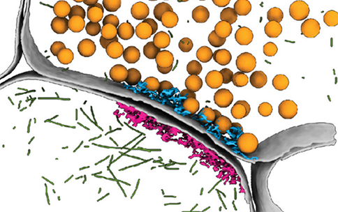
A new discovery, supported by the NIH Common Fund’s Transformative Cryo-EM Program (CryoEM), helps explain how neurons in the brain communicate with each other. The study, led by Richard G. Held, Ph.D., at Stanford University, used research tools called cryo-electron tomography (cryoET) to study synapses—the small gaps between neurons where they pass information to each other—in extraordinary detail.
Synapses have previously been too difficult to study at the molecular level due to their small size, less than one millionth of a meter in some cases. Using advanced tools provided by the Stanford-SLAC Cryo-ET Specimen Preparation Center (SCSC), researchers were able to obtain three-dimensional images of synapses at a very small scale to understand how they are organized in mouse brains. Visualizing the miniscule synapses in such detail required sample preparation with a technique called focused ion beam milling to carefully shave off unwanted layers of the frozen samples with a precise beam of ions, allowing a clearer image of synapses and associated proteins in the electron microscope. The findings, published in the Proceedings of the National Academy of Sciences, revealed that the shape of the synapse is important for neurons to transmit signals, and that the geometric properties of synapses may play an important role in neuron communication.
The NIH Common Fund’s Transformative Cryo-EM Program enables scientists to use advanced cryogenic electron microscopy (cryo-EM) and cryo-ET technology to study biological structures as close to their physiological setting as possible to learn more about the basis of health and disease of biological systems like synapse function. This new insight into synaptic organization highlights the complexity of how neurons communicate and may ultimately help researchers better understand how the brain works and how certain brain disorders like Alzheimer's or Parkinson's develop.
Nanoscale architecture of synaptic vesicles and scaffolding complexes revealed by cryo-electron tomography. Held RG, Liang J, Brunger AT. Proc Natl Acad Sci U S A. 2024 Jul 2;121(27):e2403136121. doi: 10.1073/pnas.2403136121. Epub 2024 Jun 26. PMID: 38923992; PMCID: PMC11228483.


