Understanding the form and function of biological molecules expands our knowledge of how living things work. Researchers can apply that knowledge to improve health, like in the process of developing new antibiotics. To answer questions about the form and function of biological molecules, many structural biologists are moving from traditional X-ray crystallography methods to cryoelectron microscopy (cryoEM). Images obtained through cryoEM methods provide similar amounts of detail as those of X-ray crystallography, allow for more protein variability than X-ray crystallography methods, and eliminate the need to find crystallization conditions. The Common Fund’s Transformative High-Resolution Cryoelectron Microscopy (CryoEM) program is broadening access to cryoEM for biomedical researchers. One way to reach this goal is by training researchers so that they may independently conduct cryoEM research projects. Dr. Gerald Jogl, a laboratory head at Brown University, studies antibiotic resistance in bacterial ribosomes using structural biology techniques such as X-ray crystallography. Ribosomes are particles in the cell that read the genetic blueprints encoded in RNA to make proteins. Many antibiotics work by targeting bacterial ribosomes so that they can no longer make proteins critical for bacterial cell survival. Antibiotic resistance can occur when the structure of a bacterium’s ribosome mutates to differ from the normal structure, making it harder for the antibiotic to recognize and target it. Dr. Jogl aims to use cryoEM to obtain images of the abnormal ribosome structures, including those not suitable for X-ray crystallography, to help advance understanding of antibiotic resistance.
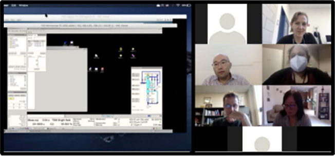
To gain the necessary expertise to conduct cryoEM studies, Dr. Jogl applied to the cross-training program offered at the Common Fund-supported National Center for CryoEM Access and Training (NCCAT). The intensive training program consists of instruction from NCCAT staff, access to equipment for sample preparation, and access to microscopes for screening and data collection, all of which was performed remotely due to the pandemic. An advantage of the remote training format was the increased flexibility for distributing and scheduling the lessons on cryoEM theory, allowing Dr. Jogl to better prepare for, review, and absorb the information. Remote demonstrations of preparing the grids that hold the sample and using the specialized cryoEM microscopes still allowed Dr. Jogl to achieve most of the training goals. Once institutional policies allow for post-pandemic in-person interactions, Dr. Jogl plans to meet the final training goals addressing practical equipment use. Dr. Jogl’s experience is one example of how the CryoEM program is broadening access to high-resolution cryoelectron microscopy through cross-training, even as the pandemic limits on-site access for biomedical researchers.


